
Articles
- Page Path
- HOME > J Powder Mater > Volume 29(2); 2022 > Article
-
Review Paper
선택적 레이저 용융법으로 제조된 CoCrFeMnNi계 고엔트로피합금의 미세조직 및 기계적 물성 연구 동향 - 박정 민*
- Microstructure and Mechanical Properties of CoCrFeMnNi-type High-entropy Alloy Fabricated by Selective Laser Melting: A Review
- Jeong Min Park*
-
Journal of Korean Powder Metallurgy Institute 2022;29(2):132-151.
DOI: https://doi.org/10.4150/KPMI.2022.29.2.132
Published online: March 31, 2022
한국재료연구원 분말재료연구본부 3D프린팅재료연구실
Department of 3D Printing Materials, Korea Institute of Materials Science (KIMS), Changwon 51508, Republic of Korea
- *Corresponding Author: Jeong Min Park, TEL: +82-55-280-3502, FAX: +82-55-280-3502, E-mail: jmpark@kims.re.kr
- - 박정민: 선임연구원
• Received: April 19, 2022 • Revised: April 22, 2022 • Accepted: April 28, 2022
© The Korean Powder Metallurgy Institute. All rights reserved.
- 3,134 Views
- 43 Download
- 7 Crossref
Abstract
- The CoCrFeMnNi high-entropy alloy (HEA), which is the most widely known HEA with a single facecentered cubic structure, has attracted significant academic attention over the past decade owing to its outstanding multifunctional performance. Recent studies have suggested that CoCrFeMnNi-type HEAs exhibit excellent printability for selective laser melting (SLM) under a wide range of process conditions. Moreover, it has been suggested that SLM can not only provide great topological freedom of design but also exhibit excellent mechanical properties by overcoming the strength–ductility trade-off via producing a hierarchical heterogeneous microstructure. In this regard, the SLM-processed CoCrFeMnNi HEA has been extensively studied to comprehensively understand the mechanisms of microstructural evolution and resulting changes in mechanical properties. In this review, recent studies on CoCrFeMnNi-type HEAs produced using SLM are discussed with respect to process-induced microstructural evolution and the relationship between hierarchical heterogeneous microstructure and mechanical properties.
- 최근 인류의 활동반경이 심해, 극지방, 우주 영역까지 빠르게 확장됨에 따라 더욱 강하고 질긴 고성능의 금속 소재에 대한 요구는 더욱 빠르게 증가하고 있다. 이를 위 해 다수의 재료공학자들은 금속 소재의 내재적인 물성 향 상을 위해 새로운 조성의 합금을 설계하거나 이로운 미세 조직을 발현시키기 위한 기법을 연구한다. 이때, 최근 재 료공학자들은 차세대 극한환경용 소재로 주목받는 고엔트 로피합금(High-entropy alloy)에 집중한다. 고엔트로피합금 은 1가지 혹은 2가지 주요 원소를 갖는 전통적인 합금계 와 달리 다수의 주요원소를 지니며, 이러한 조성적 특수성 으로 인해 우수한 기계적 물성을 나타내는 것으로 알려지 고 있다[1-3]. 특히, 서로 다른 원자 반경을 지닌 주요원소 들은 고엔트로피합금 내부 극심한 격자뒤틀림(severe lattice distortion)을 야기하게 되고, 이를 기반으로 한 높은 고용강화 효과는 합금의 항복강도 향상뿐만 아니라 우수 한 가공경화율 및 고연신율에도 영향을 미치는 것으로 알 려져 있다[3-5]. 또한, 제한적인 조성계에서 특정 원소 함 량을 변화시키며 물성 변화를 탐구하는 기존의 합금설계 방식과 비교하여, 다중주요원소합금(Multi-principal element alloy)의 개념을 가진 고엔트로피합금은 조합 가능한 원소 종류 및 비율이 무궁무진함에 따라 금속 소재의 성능 향 상을 위한 합금설계 방법론을 더욱 넓은 범위로 확장시켰 다는 점에서 큰 의의를 가진다[3-5]. 이에 따라 지난 15년 간 전세계 금속 학계에서는 우수한 기계적 물성과 다기능 성을 지닌 다양한 조성의 고엔트로피합금들이 폭발적인 속도로 개발되어 왔으며, 그 연구 속도 및 관심도는 지금 도 더욱 가파르게 증가하고 있다[5-10].
- 한편, 현재까지 보고된 다양한 조성의 고엔트로피합금 들 중 가장 많은 연구가 이루어진 합금은 2004년 영국 Oxford 대학의 Brian Cantor 교수에 의해 처음 보고된 CoCrFeMnNi 고엔트로피합금(i.e., Cantor alloy)이다[11]. 해당 합금은 서로 다른 5개의 원소가 동분율로 혼입되어 있음에도 넓은 온도 영역에서 안정한 FCC 단상을 나타내 며 우수한 극저온 인장강도-연신율 조합[11-13], 파괴인성 [13], 내식성[14], 내수소취성[15, 16], 관통저항성[17] 등을 보임에 따라 전세계적으로 많은 관심을 받으며 다양한 연 구들이 이루어져 왔다. 특히, 해당 합금은 세계 최초로 보 고된 고엔트로피합금 중 하나임에 따라 신합금 설계의 모 합금(reference alloy)으로서 활용되며, 원소간 비율을 제어 한 비등원자 CoCrFeMnNi 합금[18], 특정 원소를 제거한 CoCrFeNi[19], CoFeMnNi 합금[20], 특정 원소를 첨가한 CoCrFeMnNiC[21], AlCoCrFeMnNi[22] 합금 등 다양한 조성의 합금들이 개발되어져 왔다. 한편, 이러한 합금들은 대부분 진공유도용해(Vacuum indction melting), 아크용해 (Arc melting) 등 주조 방식을 통해 제작되어 연구되어 왔 다[11-22]. 그러나 주조로 제작된 고엔트로피합금은 조대 한 결정립과 주조 결함 등을 내포함에 따라, 압연, 단조 등 의 소성가공으로 주조 조직을 파괴하고 후열처리를 통해 균질한 등축정의 결정립을 얻는 후공정의 적용이 많이 활 용되어져 왔다[12-18]. 특히, FCC 단상의 CoCrFeMnNi계 합금은 매우 우수한 인장 연신율을 나타내는 것과 대비해 다소 낮은 항복강도를 지님에 따라, 해당 합금계의 항복강 도를 향상시키기 위한 부분재결정 활용[23], 초미세립화 [24], 불균질 결정립화[25] 등 복잡한 후공정을 적용하는 연구들이 계속해서 이루어져왔다. 그러나, 이러한 기법들 은 대부분 고엔트로피합금 최종재의 크기 및 형상에 제약 을 주는 단점이 존재하며, 이러한 기법들로 향상시킬 수 있는 해당 합금계의 강도와 연신율 조합은 점차 한계점에 도달하고 있다.
- 이때, 최근 고엔트로피합금 학계에서는 해당 합금계에 대한 금속 적층제조(Metal additive manufacturing) 기술 적용에 주목한다[26-28]. 금속 3D 프린팅 기술로도 불리어 지는 금속 적층제조 기술은 CAD data로 설계된 3차원 형 상의 제품을 한 층씩 쌓아 올리며 제작하는 Bottom-up 방 식의 공정 특성을 나타내며, 높은 설계자유도를 기반으로 전통 공법으로 제조하기 불가능한 고성능/다기능성의 복 잡한 형상의 제품을 한번에 제작할 수 있는 장점을 지녀 기존 부품 제조 산업의 패러다임을 바꿀 혁신적인 공법으 로 주목받는다[27-29]. 그중, 가장 대표적인 적층제조 기술 인 선택적 레이저 용융법(Selective laser melting, SLM)은 그림 1과 같이 수십 마이크로미터 수준의 미세한 구형 금 속 분말을 베드에 균질하게 도포하고 고밀도 레이저빔을 선택적으로 조사하여 분말을 용융/응고시키는 작업을 높 이 방향(적층 방향)으로 반복하며 3차원 형상의 제품을 제 작하는 공법이다[29-31]. 즉, 해당 기술은 직접 에너지 증 착법(Directed energy deposition) 등 타 적층제조 공정 대 비 우수한 자유도와 정밀도를 지닌 것으로 알려짐에 따라 항공우주, 모빌리티, 의료용 임플란트 산업 등 복잡한 형 상의 부품을 요구하는 산업에서 가장 널리 활용되어지고 있는 적층 제조 기술로 알려지고 있다[29-31].
- 이때, 최근 보고에서는 SLM 공정이 적용된 소재가 계 층적이며 불균질한 미세조직을 나타내며 이러한 미세조직 학적 양상이 상용 공법 적용 소재 대비 항복강도와 연신 율 조합을 비약적으로 향상시킬 수 있다는 사례가 등장하 였다[32, 33]. 이에 따라, 기존의 공정 최적화 및 형상 설 계 연구가 주로 이루어지던 전세계 적층제조 학계에서는 SLM 공법 적용을 통한 소재들의 미세조직 제어 및 물성 향상 연구에 대한 관심이 폭발적으로 증가하였으며, 최근 5년간 다양한 소재들에 대한 공정-미세조직-기계적 물성 상관 관계 연구들이 활발히 이루어져 왔다[27-33]. 이때, CoCrFeMnNi계 합금은 SLM 공정이 적용 가능한 소재들 중 넓은 공정 조건에서 안정한 단상의 건전한 시편이 제 조 가능함에 따라 해당 합금을 기초 소재로 한 공정-미세 조직-물성 연구들이 활발히 이루어져 왔으며, SLM 공정 제어 및 첨가 원소를 통해 해당 합금계의 기계적 물성의 한계치를 넘기 위한 시도들이 다수 이루어져 왔다[34-38]. 즉, 고성능 구조재료인 고엔트로피합금에 대한 SLM 공정 적용은 적층제조 학계에서의 소재적인 측면의 기초 연구 활용뿐만 아니라 우수한 기계적 물성을 부여할 수 있는 방법론, 해당 합금계 기반의 첨단 부품 제조 가능성을 획 기적으로 앞당길 수 있는 접근법으로써 간주되며, 이에 대 한 연구 관심도는 금속학계에서 매년 더욱 가파르게 증가 하고 있다. 본 리뷰 논문에서는 대표적인 CoCrFeMnNi계 고엔트로피합금들에 대한 SLM 공정 적용 사례들과 조형 체의 미세조직학적 특성 및 기계적 거동 분석 관련 최근 연구 결과들을 설명함으로써 국내외 관련 학계의 연구에 도움을 주고자 한다.
1. 서 론
Fig. 1
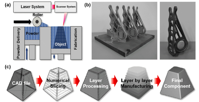
(a) A schematic of SLM process. (b) SLM-produced brackets (left) on a build plate and finished part (right) for Airbus A350 with topology optimized design resulting in ~30% weight reduction of conventional part (reprinted from [30] with permission from Elsevier). (c) Schematic illustrations indicating the process of tool path generation from CAD data and layer processing for additive manufacturing [29].
- 2.1. SLM 조형체의 미세조직학적 특성
- CoCrFeMnNi 합금은 높은 내식성과 더불어 넓은 온도 범위에서 안정한 FCC 상을 형성함에 따라 취성의 이차상 및 준안정상의 출현으로 인한 크랙 형성 등이 방지되며 상대밀도 99.9% 이상의 건전한 시편이 제조 가능한 우수 한 SLM 공정 적용성(Printability)을 나타내는 것으로 보고 되고 있다[34]. 특히, CoCrFeMnNi 고엔트로피합금은 SLM 공정 도중 상변태가 일어나는 Ti 합금, 석출상이 나타나는 Al 및 Ni 합금, 고반사율로 인한 난적층성의 Cu 합금과 달 리 넓은 공정 조건에서 안정한 FCC 단상의 건전한 미세 조직을 나타냄에 따라, 아직까지 완벽하게 구축되지 않은 SLM 조형체의 공정-미세조직-물성 상관관계에 대한 원천 연구를 위한 기초 소재로써 활용되어진다[34]. 영국 Imperial College의 Pham 그룹은 그림 2와 같이 SLM 공정 도중 금 속 소재의 미세조직 성장에 대한 기초 연구를 위해 CoCrFeMnNi 합금을 기초 소재로 활용하여 다양한 연구 를 수행하였다[34-36]. 그들은 구형의 CoCrFeMnNi 합금 분말을 기반으로 SLM 장비인 Renishaw AM250 장비를 활용해 다양한 공정 조건에서 상대밀도 99.5% 이상의 건 전한 CoCrFeMnNi 합금 조형체를 제작하였다. 그들은 그 림 2(a-c)와 같이 CoCrFeMnNi 합금의 용융풀 경계부의 열 흐름에 따른 결정립 성장 방위와 결정립들의 적층 방 향으로의 Epitaxial 성장 경향 간의 상관관계를 탐구하며, SLM으로 제조되는 FCC 구조 금속소재의 극심한 결정학 적 t extu re와 불균질한 결정립 형상의 근원을 탐구하였다 [34-36]. 또한, 그들은 그림 2(d, e)와 같이 CoCrFeMnNi 합금의 용융풀에서의 결정립 성장 양상에 대한 이해를 바 탕으로 SLM 공정 시 서로 다른 스캔 패턴과 스캔 방향이 적용된 FCC 구조 금속소재의 결정립들의 형상 및 배열의 변화와 기계적 물성 변화에 대한 건설적인 가이드라인을 제시하였다[35].
- 한편, 그림 3과 같이 SLM으로 제조된 CoCrFeMnNi 합 금은 앞서 기술한 용융풀(Fig. 3(a)), 불균질한 결정립 형 상 및 크기 분포(Fig. 3(b)), 극심한 결정학적 텍스처(Fig. 3(d))뿐만 아니라, 높은 분율의 저경각입계(Fig. 3(e))와 높 은 전위밀도(Fig. 3(f))와 상관있는 나노 전위셀 구조(Fig. 3(c, g, h) 등 수십 나노미터에서 수백 마이크로미터 단위 까지 계층적이며 불균질한 미세조직을 함유하는 것으로 보고되고 있다[37]. 특히, SLM 공정을 통해 유도되는 수 백 나노미터 수준의 직경을 가지는 나노셀 구조는 소성 변형에 기인한 전위 셀 구조와는 다르게 경계부의 용질원 자 편석을 동반하는 등 고유한 특성을 지닌 것으로 보고 되고 있다[32, 33, 39].
- 즉, SLM 공정 도중 초고속의 냉각 속도(106 - 108 K/s) 를 동반하는 가혹한 열이력은 조형체 내부 높은 열 응력 을 야기하게 되고 이는 다량의 전위를 생성시켜 셀 구조 의 전위 네트워크를 야기하게 된다[39]. 이러한 나노 전위 셀 조직은 그림 4(a-d)와 같이 용융풀 내의 열 구배 및 열 흐름 양상 차이에 따라 성장 방향과 크기가 달라지며, 특 히 그림 4(b, d)에서 나타나는 것과 같이 열 흐름의 반대 방향이자 기존에 성장한 결정립과 Epitaxy를 최대한 유지 할 수 있는 방향으로 육각형의 단면을 가진 실린더 형태 로 자라남에 따라 관측 위치에 따라 육각형 모양, 기둥 모 양 등 그 형상 및 분포가 달라진다[39, 40]. 또한, 셀 조직 의 형성 시 열역학적 요인인 조성적 과냉, 속도론적인 측 면의 Bernard Marangoni 표면 불안정성 등으로 인해 셀 경계부에는 용질 원자의 편석을 함께 동반하는 것으로 보 고되고 있다[39]. 이때, 일부 합금계에서는 이러한 용질 원 자 편석은 SLM 조형체의 셀 경계부의 전위이동 방해능을 더욱 향상시켜 변형 기반의 셀 조직 대비 적층 제조 기반 의 셀 조직이 기계적 물성에 더욱 유리하다고 보고되고 있다[32]. 그러나, 이러한 편석 기반 셀 경계 강화효과의 정도에 대해서는 아직까지 의견이 분분하며 최근 논문들 에서는 셀 경계부의 기계적 물성 기여도는 일반적인 전위 밀도 기반 강화효과와 그 크기가 흡사하다는 의견들도 제 기되어 지고 있다[37,39]. 즉, 편석에 의한 셀 경계 강화 효과는 편석 되는 원소 종류와 기지상의 조성에 따라 그 영향이 달라질 수 있음에 따라, 향후 합금들 및 구성원소 에 따른 편석 기반 셀 경계 강화 효과를 정량화할 수 있 는 연구가 필요할 것으로 판단된다. 한편, CoCrFeMnNi 합금은 그림 4(e)의 열역학 계산 결과와 같이 응고 시 Mn 과 Ni원자가 응고선단으로 배출되면서 주조재, 용접재의 수지상간영역(Interdendritic region)에서는 Mn과 Ni 성분 의 편석이 자주 관측된다[40]. 이와 유사하게 SLM으로 제 조된 CoCrFeMnNi 합금 조형체의 셀 경계부는 그림 4(fl) 와 같이 Mn 및 Ni의 편석이 관측되는 것으로 보고되고 있다[40]. 또한, 셀 경계부에는 그림 4(h, l)과 같이 Mn 성 분의 편석과 동시에 주로 MnO 혹은 MnS 등의 산화물, 황화물이 수십 나노미터 크기의 석출물의 형태로 분포하 며 이러한 나노 석출물들은 조형체의 기계적 물성에 직접 적인 영향을 끼치는 것으로 보고되고 있다[40-42].
- 즉, CoCrFeMnNi 합금에 대한 SLM 공정의 적용은 물성 향상을 위해 기존에 제어 가능하였던 제한적인 미세조직 학적 변수에 SLM 조형체 고유 미세조직인 나노 석출물, 나노 셀조직, 불균질 결정립 분포, 용융풀 등으로 확장시 킬 수 있는 것에 의의가 있으며, 이를 유용하게 활용할 시 복잡한 공정 없이 한번의 공정 조건 변화만으로 소재의 나노 단위부터 거시적인 부품 형상까지 고려할 수 있는 맞춤형 물성의 부품 설계가 가능할 것으로 이해할 수 있 다. 그러나, SLM 조형체의 계층적 불균질 미세조직이 소 재 변형 단계별 미치는 영향에 대한 이해는 아직까지 명 확하게 구축되지 않은 형태임에 따라, 최근 학계에서는 CoCrFeMnNi 조형체의 다양한 미세조직학적 인자들이 기 계적 물성에 미치는 기여도에 대한 정량화 연구가 폭발적 으로 이루어지고 있다.
- 2.2. 미세조직-기계적 물성 상관관계 연구동향
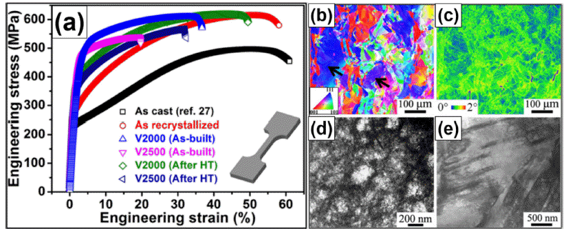
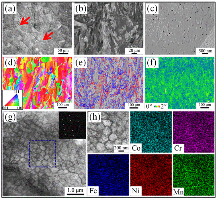
- 싱가폴 SIMTech의 Nai 그룹[37]은 가스분사법(Gas atomization)으로 제조된 구형의 CoCrFeMnNi 합금 분말 을 활용해 3D systems 社의 SLM 장비인 ProX300 장비 기반 상대밀도 99% 이상의 고밀도 조형체를 제작하였고, 그림 5(a)와 같이 해당 조형체가 주조재 및 재결정재 대비 우수한 항복강도-연신율 조합을 나타냄을 보고하였다. 특 히, 그들은 그림 5(b, c, e)와 같이 SLM 조형체의 변형 도 중 변형쌍정의 출현에 따른 우수한 연신율과 더불어, 그림 5(d)와 같이 조형체의 계층적 불균질 미세조직 인자들 중 나노 전위셀 구조가 조형체의 높은 항복강도에 가장 지배 적으로 기여하는 것으로 보고하였다.
- 즉, SLM 공법을 통해 유도된 수백 나노미터 크기의 셀 조직은 기존 공법으로 제조된 CoCrFeMnNi 합금과 핵심 적인 미세조직학적 차이임과 동시에 기계적 물성 향상에 직접적으로 영향을 끼침에 따라, 다수의 학자들은 이러한 셀 구조의 기계적 물성 기여도에 대한 정량화 연구와 이 를 활용한 물성 향상 연구들을 주로 진행해 왔다. Kim 등 [41]은 GE 社의 소형 SLM 장비인 Mlab 장비로 제조된 CoCrFeMnNi 합금의 셀 경계부의 높은 전위밀도 뿐만 아 니라 나노 산화물도 변형 도중 전위의 이동을 저지해주어 기계적 물성 향상에 기여할 수 있다고 보고하였다. 또한, 그들은 그림 6(a-d)의 TEM 이미지와 같이 나노 전위셀 경 계부의 나노 산화물은 기존 공법으로 제조된 CoCrFeMnNi 합금 대비 SLM 조형체의 인장물성 뿐만 아니라 고주기 피로특성(그림 6(e)) 및 크립 특성(그림 6(f))도 향상시킬 수 있다고 보고하였다[41-43].
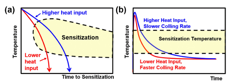
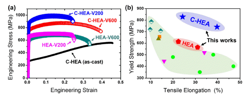
- Park 등[44]은 이러한 셀 경계부를 더욱 직접적으로 강화 시키기 위해 치환형 원소로만 구성되어 있는 CoCrFeMnNi 고엔트로피합금에 소량의 탄소를 첨가한 SLM 조형체를 제작하였다. 그들은 진공유도용해(Vacuum induction melting) 을 통해 탄소를 1 at% 첨가한 CoCrFeMnNi 합금과 탄소 를 첨가하지 않은 CoCrFeMnNi 합금 잉곳을 제조하였고, 해당 잉곳들을 기반으로 가스분사법을 통해 합금화된 구 형의 분말을 제작하여, 최종적으로 GE 社 Mlab 장비 기 반 상대밀도 99% 이상의 SLM 조형체들을 제작하였다. 이때, 그들은 그림 7(a)와 같이 탄소를 1 at% 함유한 고엔 트로피합금 조형체들이 탄소를 함유하지 않은 조형체 대 비 항복강도가 200 MPa 이상 향상되며 연신율도 동시에 증가함을 확인하였으며, 그림 7(b)와 같이 기존에 보고된 CoCrFeMnNi계 합금 조형체 대비 월등히 우수한 강도-연 신율 조합을 보임을 확인하였다. 이러한 탄소 첨가 고엔트 로피합금 조형체의 월등한 기계적 물성 향상은 그림 8와 같이 SLM 조형체의 계층적 불균질 미세조직인자들 중 불 균질한 결정립 형상 및 크기분포(그림 8(a)), 높은 잔류 전 위밀도(그림 8(b))에 관계된 나노셀 구조(그림 8(c)) 등과 더불어 셀 경계부에 석출된 수십 나노미터 크기의 경한 나노탄화물이 핵심 인자인 것으로 보고되어진다. 즉, SLM 공정 도중의 반복적인 입열은 탄소 함유 CoCrFeMnNi 조 형체가 Cr23C6 탄화물을 형성하기 용이한 예민화온도에 반복적으로 노출되게 만들어 주며, 해당 온도 영역대에서 탄소와의 친화성이 좋은 Cr 성분이 셀 경계부로 빠르게 확산됨에 따라 수십 나노미터단위의 탄화물이 결정립 내 부 셀경계에 균질하게 분포할 수 있게 된다. 이를 통해 SLM 조형체는 탄소 첨가에 따라 주로 결정립계에 조대한 탄화물 석출을 동반해 소폭의 강도 상승과 급격한 연신율 의 감소를 보여주는 기존 공법 제작제와 달리 균질하게 분포된 나노탄화물을 기반으로 탄소 첨가를 통한 강화효 과를 극대화시킬 수 있는 것을 확인할 수 있다[21, 44].
- 특히, 이러한 셀 경계부 나노 탄화물은 전위의 이동을 방해해 그림 9와 같이 셀 경계부의 다량의 전위 집적 (Dislocation pile-up)을 야기하게 된다. 즉, 그림 9(a, b)의 조형체의 변형 전 상태와 비교하여, 그림 9(c-f)와 같이 변 형량의 증가에 따라 다량의 전위가 셀경계부 및 나노 탄 화물에 집적되게 되고, 이는 조형체의 소성 변형 도중 높 은 역응력(Back stress)을 야기하게 된다. 이러한 높은 역 응력은 조형체의 변형 도중 가공경화율을 높여 넥킹 등의 소성 불안정성을 지연시키고, 최종적으로 탄소 함유 고엔 트로피합금이 탄소를 함유하지 않은 고엔트로피합금 조형 체 대비 더욱 향상된 균일 연신율(Uniform elongation)을 갖는 것에 기여하게 된다[45, 46]. 이는 통상 탄소 첨가에 따라 강도는 증가하지만 연신율은 감소하는 기존 공법 적 용재와 명확히 대비된다.
- 한편, 그림 7(a)에서는 탄소 함유 고엔트로피합금은 SLM 공정 시 스캔 속도의 변화(200 mm/s vs. 600 mm/s) 에 따라 탄소 함유 고엔트로피합금 조형체들은 탄소를 함 유하지 않은 조형체과 대비하여 더욱 급격한 기계적 물성 의 급격한 변화를 나타내는 것을 확인 가능하다[44]. 해당 양상은 그림 10와 같이 공정 도중 시편에 가해진 입열량 차이에 따른 예민화 온도 노출 시간 차이로 인한 탄화물 분율 변화에 기인하는 것으로 보고되고 있다[44]. 이는 SLM 조형체 내부 나노셀 경계 특성을 첨가 원소 분율 조 절뿐만 아니라 동일 조성 내에서의 공정 조건의 변화를 통해서도 충분히 제어할 수 있다는 것을 보여준다.
- 이와 흡사하게 Chen 등[47]은 최근 볼 밀링 공정을 활 용하여 구형의 CoCrFeMnNi 분말에 50 nm 크기의 나노 TiC 입자를 부착한 복합재 분말을 그림 11(a, b)와 같이 제 조하였으며, 해당 분말에 대한 SLM 공정을 적용해 조형 체를 제작하고 순수 CoCrFeMnNi 합금 조형체와 미세조 직 및 기계적 물성을 비교 분석하였다. 그들은 그림 12(ad) 와 같이 SLM 공정 도중 TiC 입자는 용융되어 Ti 성분 과 C 성분으로 분리되고, 해당 성분들은 응고 시 조형체 내부 나노 셀 경계부에 수십 나노미터 크기의 TiC1-x 타입 의 탄화물로써 석출되는 것을 확인하였다. 그들은 이러한 나노셀 강화 효과를 통해 그림 12(e)와 같이 TiC 첨가 CoCrFeMnNi 합금 조형체가 순수한 CoCrFeMnNi 합금 조형체 대비 300MPa 수준의 높은 인장강도 향상과 연신 율의 향상이 이뤄질 수 있음을 보고하였다.
- 즉, SLM 공정에 기인한 특유 미세조직인 나노 셀 구조 의 활용은 통상의 금속소재가 나타내는 강도-연신율 역전 현상을 극복하고 기계적 물성을 획기적으로 향상시킬 수 있는 방법론으로써 충분히 사용될 수 있으며, 다양한 고엔 트로피합금들에 대한 첨가 원소 및 공정 제어 기반의 나 노셀 크기 제어, 경계부 편석 제어 및 석출상 제어 관련 연구들은 현재 진행형으로 활발히 진행되고 있다[48-52].
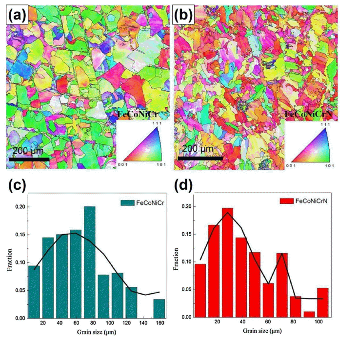
- SLM으로 제조된 CoCrFeMnNi 합금의 계층적 불균질 미세조직학적 인자들 중 나노 셀 구조의 높은 기계적 물 성 기여도와 더불어 불균질한 결정립 형상과 크기 분포도 조형체의 우수한 기계적 물성에 기여하는 것으로 보고되 고 있다[53]. 즉, 균질한 결정립 크기 분포를 가지는 소재 와 달리 SLM 조형체 내부 불균질한 결정립 크기 분포는 변형 도중 높은 변형률 구배(Strain gradient)를 발생시켜 다량의 기하학적 필수 전위(Geometrically necessary dislocation) 증식을 야기하게 되고 이는 불균질 변형 강화효과 (Hetero-deformation-induced hardening)를 야기시켜 조형 체의 가공경화율을 향상시키는 데 기여할 수 있다[53, 54]. Song 등[54]은 질소 분위기에서 가스분사법을 활용해 질 소가 첨가된 CoCrFeNi 고엔트로피합금 분말을 제조하였 고, 해당 분말 기반의 SLM 조형체를 제작하였다. 그들의 결과에서는 그림 13과 같이 질소를 첨가한 CoCrFeNi 합 금 조형체가 질소를 함유하지 않은 경우와 비교하여 더욱 불균질한 결정립 크기 분포를 나타내는 것으로 보고되어 진다. 또한, 그들은 질소 첨가 조형체의 불균질한 결정립 구조가 조형체의 변형 도중 높은 역응력을 야기시켜, 그림 14와 같이 질소를 함유하지 않은 조형체 대비 높은 강도 레벨과 동시에 향상된 가공경화율 및 향상된 균일 연신율 을 유도할 수 있음을 보고하였다[54].
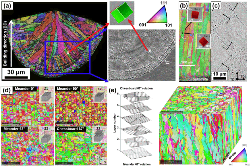
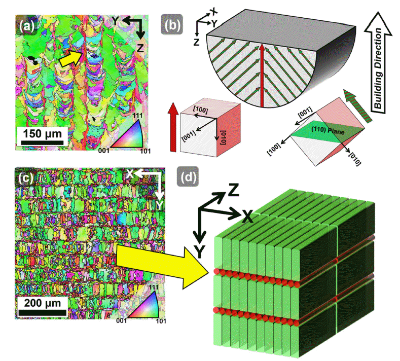
- 한편, SLM 조형체의 결정립 형상 및 크기 분포의 공간 불균질성(Spatial heterogeneity)는 스캔 패턴 및 적층 방향 에 따라 크게 달라진다[35, 36]. 우선 SLM 조형체 내부 결 정립들은 응고를 통해 성장함에 따라 극심한 결정학적 텍 스처를 나타냄과 동시에 적층방향으로 연신된 주상정의 결정립 형상을 갖게 된다. 예를 들어 CoCrFeMnNi 분말에 대한 지그재그 형태의 레이저 스캔을 층별로 180도 회전 시키며 SLM 공정을 수행할 경우, 그림 15(a, b)와 같이 용 융풀의 중심부에는 적층 방향으로 응고 시 우선성장방위 인 <100>의 결정방위를 갖는 실린더 형태의 결정립이 자 라나지만, 용융풀의 측면부에는 적층 방향과 45도의 각도 를 가진 <100> 방위 결정립들이 자라남에 따라 적층방향 으로 <110>의 결정 방위를 지닌 결정립이 나타난다[35, 53]. 특히, 레이저가 반복해서 지나가며 각 용융풀 측면부 는 서로 오버랩 됨에 따라 높은 입열로 인해 적층 방향으 로 <110> 텍스처를 가진 조대한 판형의 결정립이 형성되 게 된다. 이러한 결정립 성장 양상은 그림 12(c, d)와 같이 적층면을 바라보았을 때 미세한 <100> 텍스처 결정립들 과 조대한 <110> 텍스처 결정립들이 스캔 방향에 평행한 리본 형태로 층층히 배열된 구조를 야기하게 된다[35, 53].
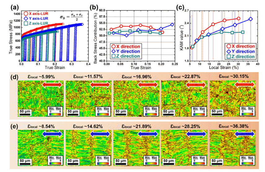
- 이러한 SLM 공정으로 유도된 결정학적 텍스처와 형상 학적 텍스처(Morphological texture)는 SLM 조형체의 기 계적 거동에 직접적인 영향을 끼친다. Park 등[53]은 서로 다른 적층 방향으로 제조된 1%C-CoCrFeMnNi 조형체에 대한 미세조직 분석과 방향별 인장변형 거동을 해석하였 다. 그들은 불균질한 결정립 크기 분포가 불균질 변형 강 화 효과를 유발하며 항복강도 및 변형 초기 가공경화능에 직접적인 영향을 끼침을 규명하였으며, 인장방향별로 주 상정 내의 전위 평균 자유 경로(dislocation mean free path)가 달라짐에 따라 형상학적 텍스처에 의한 기계적 이 방성이 충분히 야기할 수 있음을 보여주었다. 해당 연구에 서는 그림 16에서 확인되는 것과 같이 미세 결정립과 조 대 결정립 리본이 등변형(iso-strain) 상태에 놓일 수 있는 X 방향(그림 15(c,d))이 변형 초기 높은 역응력으로 인해 (그림 16(b)) 높은 항복강도를 보임을 확인하였다. 특히, 변형 도중 미세 결정립과 조대 결정립 간의 극심한 변형 구배에 따라, 그림16(c-d)와 같이 미세결정립과 조대결정 립간의 계면에서 기하학적 필수 전위가 활발히 증식되게 되고 이는 변형 초기 불균질 변형 강화 효과가 극대화될 수 있는 X 방향에서 높은 가공경화율과 인장강도에 기여 하게 된다[53]. 또한, 조형체 내부 극심한 결정학적 텍스처 는 상대적으로 낮은 적층 결함 에너지(Stacking fault energy) 를 갖는 CoCrFeMnNi 합금의 인장방향별로 변형 쌍정 (Deformation twin) 거동의 차이를 야기해 변형 후반부의 가공경화 거동 및 인장강도, 연신율에 영향을 끼침이 보고 되었다.
- Jin 등[55]은 그림 17과 같이 각각 서로 다른 스캔패턴 으로 조형된 CoCrFeMnNi 합금에 대한 피로 특성을 분석 하였고, 스캔 패턴의 변화에 따른 결정립 형상 및 배열 변 화에 따라 인장 물성뿐만 아니라 피로 저항성도 급격히 달라짐을 확인하였다. 해당 연구에서는 층간 회전 없이 지 그재그의 레이저 경로를 가지는 Meander 패턴으로 조형 한 시편이 가장 단축이 좁은 주상정 결정립 배열을 가짐 에 따라, 크랙 성장 방향인 주상정의 단축 방향으로 다량 의 결정립계가 크랙의 성장을 저지시킴에 따라 가장 높은 피로 저항성을 나타내는 것으로 보고되었다.
- 즉, SLM 공정을 통해 원하는 용도에 맞는 부품 제조를 위해선 공정 설계 시 최적화된 스캔 방향 및 적층 방향의 설정이 필수적이며, 이를 위해선 조형체의 변형 거동에 영 향을 미치는 계층적 불균질 미세조직학적 인자들 각각의 기계적 물성 기여도를 고려한 공정 설계가 요구됨을 알 수 있다. 향후에는 이에 대한 더욱 견고한 데이터베이스들 이 구축될 것으로 전망되며, 적층제조 기반 고엔트로피합 금 조형체에 대한 변형 도중 미세조직 변화에 대한 실시 간 관측 및 해석[56-59], 인공지능(Artificial intelligence, AI) 및 기계적학습(Machine learning, ML) 기반 미세조직- 기계적 물성 상관관계 연구[60-62] 등이 활발히 진행될 것 으로 전망한다.
- 2.3. 조형체 후열처리 연구동향
- SLM으로 제조된 부품은 내부의 잔류응력을 제거하고 품질에 대한 신뢰성을 확보하기 위해 후열처리가 자주 이 루어진다. 이에 따라 상용 소재들인 스테인리스강, Ni기 초합금, Ti 합금 등은 이러한 후열처리에 따른 미세조직 및 기계적 물성 변화에 대한 연구가 계속해서 이루어져 왔으며, 이를 통해 각 소재별 최적 물성을 확보할 수 있는 후열처리 조건의 표준 설립을 노력이 전세계적으로 활발 히 이루어지고 있다. 이와 달리 SLM으로 제조된 고엔트 로피합금에 대한 후열처리 연구는 아직까지 많이 이루어 지지 않았으며 극소수의 논문들만이 발표되어 왔다. Nai 등[37]은 SLM으로 제조된 CoCrFeMnNi 조형체에 900°C 에서 1시간의 열처리를 수행하였고, 해당 조형체들은 열 처리 이후 항복강도는 낮아지나 인장강도는 거의 유지되 며 연신율이 증가함을 보여주었다. Li 등[38]은 상대밀도 98% 수준의 SLM 기반 CoCrFeMnNi 합금 조형체에 대해 1150°C의 고온에서의 열간등방가압(Hot isostatic pressure, HIP) 공정을 진행하였고, 해당 조형체는 HIP 처리 후 상 대밀도 99% 이상으로 치밀화됨과 동시에 항복강도는 소 폭 감소하지만 인장강도는 향상됨을 확인하였다. Lin 등 [63]은 SLM으로 제조된 CoCrFeNi 합금에 대해 서로 다 른 온도 조건(500°C - 1300°C)에서 2시간씩 후열처리를 진 행하고 미세조직 변화와 기계적 물성 변화에 대한 탐구를 진행하였다. 그들은 후열처리가 수행된 CoCrFeMnNi 조 형체는 그림 18(a)와 같이 기존 공법 제작재와 흡사하게 열처리 온도의 증가에 따라 강도는 감소하고 연신율은 증 가하는 양상을 보여주나, 샤르피 충격 에너지는 그림 18(b)와 같이 열처리 온도 증가에 따라 월등히 향상됨을 확인할 수 있었다. 이때, 그들은 그림 19(a-f)와 같이 SLM 으로 제조된 CoCrFeNi 합금은 비록 열처리 온도의 증가 에 따라 잔류 응력은 줄어들지만 1100°C 이하의 온도에서 는 2시간의 긴 열처리 시간에도 불구하고 여전히 높은 전 위밀도를 함유하고 재결정이 완벽히 이루어지지 않았음을 확인하였다. 이러한 양상은 냉간압연 등 기존 공법으로 제 조된 FCC단상의 CoCrFeMnNi, CoCrFeNi 고엔트로피합 금이 통상 800°C - 900°C에서 10분의 열처리에도 완전 재 결정이 일어나는 양상[17]과 확연히 대비되며, 이는 SLM 조형체 내부의 전위셀 구조가 기존공법 제작제 대비하여 더욱 안정하다는 것을 암시한다.
- Park 등[64]은 SLM으로 제조된 탄소 함유 CoCrFeMnNi 합금에 대해 각각 800°C 및 900°C에서의 후열처리를 수행 한 연구를 보고하였으며, Lin 연구팀의 보고와 달리 해당 조형체는 그림 20(a)와 같이 열처리 이후 오히려 인장강도 가 더욱 증가하는 것을 확인하였다. 그들은 이러한 탄소 함유 조형체들은 고온의 800°C - 900°C 조건에서 재결정 이 거의 일어나지 않았으며 그림 20(b)와 같이 나노 전위 셀 구조가 잔존함을 확인하였다. 이러한 양상은 셀경계부 에서의 나노 탄화물이 셀경계의 파괴를 지연시켜주는 효 과와 동시에 내부의 침입형 탄소원자들이 재결정을 지연 시킴에 따라 발생하는 것으로 보고되고 있다. 그림 20(c) 는 CALPHAD 기반 열역학 소프트웨어인 Thermo-calc로 계산된 1%C-CoCrFeMnNi의 평형상태도를 보여준다. 즉, 해당 합금은 일반적인 316L 스테인리스강 대비 높은 탄소 함량과 Cr 함량을 가짐에 따라, 더욱 넓은 온도 영역에서 Cr23C6 탄화물이 안정하게 석출됨을 확인할 수 있다. 이에 따라 열처리가 수행된 탄소 함유 CoCrFeMnNi 조형체는 열처리 전과 대비하여 더욱 높은 분율의 탄화물을 가지게 된다. 특히, 그림 20(d-h)와 같이 증가한 분율의 나노 탄화 물들은 변형 도중 셀 경계부의 전위이동방해 효과를 증가 시키며, 셀 경계에 집적된 다량의 기하학적 필수 전위들은 높은 역응력을 발생시키게 된다. 즉, 탄소 함유 고엔트로 피합금 조형체는 후열처리 이후 잔류 전위들의 존재와 탄 화물 분율 증가로 인해 소폭의 항복강도 감소만을 보여주 며, 오히려 셀 경계부에서 야기되는 역응력으로 인해 가공 경화율이 증가하며 인장강도 및 균일 연신율이 동시에 향 상되게 된다. 이러한 양상은 탄소 함유 고엔트로피합금 조 형체에 대한 후열처리는 잔류응력 감소를 통한 부품 신뢰 성 향상뿐만 아니라 기계적 물성을 향상시킬 수 있는 방 법으로써 활용될 수 있음을 보여준다.
- 최근 Wang 등[65]은 그림 21와 같이 CoCrFeMnNi 분말 과 4 at%의 TiAl 분말을 혼합한 복합분말을 제조하여 SLM 공정을 적용하고 1175oC에서의 HIP처리와 700oC에 서의 시효열처리(Aging treatment)를 수행한 연구를 보고 하였다. 그들은 이러한 접근을 통해 그림 22와 같이 조형 체 내부 Al2O3, 시그마상, L12, B2 상 등 다양한 종류의 나 노석출물들을 석출시켰다. 이때, 그림 23(a)와 같이 열처 리를 수행하지 않은 TiAl첨가 CoCrFeMnNi 조형체는 기 존에 보고되는 순수 CoCrFeMnNi 조형체와 유사한 600 MPa 수준의 항복강도 및 인장강도를 보이지만, HIP과 시 효열처리가 수행된 조형체는 다양한 나노 석출상들의 존 재로 인해 가파른 가공경화율과 더불어 1 GPa 수준의 인 장강도를 보임을 확인할 수 있다. 또한, 그림 23(a)에서 해 당 시편들의 총 연신율은 열처리 전후 큰 차이를 보이지 않지만, 그림 23(b)의 가공경화율 커브에서 진응력-진변형 률과 가공경화율이 만나는 지점에서의 연신율은 HIP+시 효열처리된 시편이 열처리전 시편 대비 1.5배 이상 높은 값을 보임을 확인할 수 있다. 즉, 본 조형체에 대한 HIP과 시효열처리는 조형체 내부 전위밀도의 감소와 더불어 다 량의 석출상을 형성시켜 석출강화 기반의 우수한 가공경 화율을 부여하고 이를 통해 인장강도와 균일 연신을 동시 에 획기적으로 향상시키는 전략으로 활용할 수 있음을 알 수 있다.
- 고엔트로피합금 조형체 관련하여 A s-bu ilt 상태가 아닌 후열처리 후 특성 변화에 관련된 연구는 학문적 중요성에 도 불구하고 아직까지 많이 이루어지지 않은 실정이며 이 는 후열처리 관련 다양한 연구들이 표준이 정립되어지고 있는 상용재들의 경우와 대비된다. 즉, 고엔트로피합금 조 형체에 대한 후열처리 연구는 학문적 관심도에 비해 아직 까지 태동기 초반인 상태임에 따라, 향후 다양한 고엔트로 피 합금들에 대한 후열처리 적용 사례 및 기계적/기능적 특성 변화에 대한 연구 논문 편수는 가파르게 증가할 것 으로 전망된다.
2. CoCrFeMnNi계 합금의 SLM 적용 연구 동향
Fig. 2

Microstructure of SLM-processed CoCrFeMnNi alloy [34-36] (reproduced with permission from Springer Nature and Elsevier): (a) EBSD-IPF map indicating solidification microstructure of a CoCrFeMnNi HEA processed by SLM in a singletrack building [34]. Note that the blue box marked in (a) was magnified as the SEM micrograph indicating solidification cells at the right side of the SEM micrograph, and a red (<001> orientation) and green (<101> orientation) colored cubic pointed by red arrow represents the crystallographic orientation of a solidification cell indicated in IPF map and SEM micrograph [35]. (b) Variation of microstructure of SLM-processed HEA from the substrate (316L stainless steel) towards the top of the sample [35]. (c) SEM image indicating the side-branching of the solidified grains between adjacent melt tracks [35]. (d) Microstructure of building plane of the samples processed by SLM in different scanning strategies [36]. (e) 3D pseudo microstructure of the CoCrFeMnNi alloy processed by 67° rotation in every layer under chessboard laser scanning patten during SLM [36].
Fig. 3

Hierarchical heterogeneous microstructure of SLM-processed CoCrFeMnNi alloy (reprinted from [37] with permission from Elsevier): (a) OM and (b) SEM images of the lateral surface of the as-built sample. (c) SEM micrograph indicating cell structure. (d) EBSD IPF map, (e) EBSD IQ map indicating high angle grain boundaries (blue lines) and low angle grain boundaries (red lines), and (f) EBSD KAM amp image of the lateral surface of the as-built sample. (g) Bright-field STEM micrograph showing cell structure, and (h) EDS mapping results for blue box indicated in (g).
Fig. 4

Solidification cell structure of SLM-processed CoCrFeMnNi alloy (reprinted from [40] with permission from Elsevier): (a) Extracted melt pool shape near the rear of the weld pool and direction of the [100]FCC primary solidification direction is expected to be mostly along to the scan direction of SLM. (b) The schematic of solidified cell structure showing honeycomb structure observed in the plane perpendicular to scan direction. (c) Extracted melt pool boundaries along the sides of the melt pools and (d) will appear like dendritic cell in the plane perpendicular to scan direction. (e) Simulated concentrations of alloying elements in liquid and solid as a function of mole fraction solid. (f) A STEM HAADF micrograph image showing cell structure with (g-l) EDS maps of the constituent elements. The two white arrows in the Mn and O maps point to two participates (MnO).
2.2.1. 나노셀 구조 효과
Fig. 5
(a) Tensile properties of SLM-processed CoCrFeMnNi alloy and post heat-treated samples (reprinted from [37] with permission from Elsevier). Deformed microstructure of the alloy produced by SLM [37]: (b) EBSD IPF, (c) EBSD KAM, and (d, e) bright-field STEM micrographs of the tensile-fractured sample.
Fig. 6

Effect of in-situ formed nano-oxide in SLM-processed CoCrFeMnNi alloy (reprinted from Refs. [42] and [43] with permission from Elsevier): (a) TEM, (b) STEM, (c) STEM-HAADF, and (d) HR-TEM micrographs showing nano-sized MnO at cell boundaries in the sample. (e) S-N curves and (f) stress dependencies of steady-state creep rate of SLM-processed CoCrFeMnNi HEAs compared with the wrought alloys.
Fig. 7

Mechanical properties of SLM-processed 1%C-CoCrFeMnNi alloy (reprinted from Ref. [44] with permission from Taylor & Francis): (a) Engineering stress-strain curves of carbon-free (labeled by HEA) and carbon-doped CoCrFeMnNi HEAs (labeled by C-HEA) fabricated by SLM compared with that of the as-cast C-HEA. Note that the laser scanning speeds of 200 mm/s and 600 mm/s used for sample building were indicated by V200 and V600 in the sample name. (b) Ashby plot of the yield strength and tensile elongation for the SLM-processed C-HEA compared with other SLM-processed CoCrFeMnNi-type HEAs.
Fig. 8

Hierarchical heterogeneous microstructure of carbon-doped CoCrFeMnNi alloy fabricated by SLM (reproduced from Ref. [44] with permission from Taylor & Francis): (a) 3D EBSD IPF maps, (b) EBSD KAM map, (c) SEM BSE micrograph, STEM micrograph showing (d) cell structure with (e) nano-precipitates, and (f) EELS mapping results for the nano-precipitate at cell boundaries in the SLM-processed alloy.
Fig. 9

Deformed microstructure of the carbon-doped CoCrFeMnNi HEA processed by SLM (reprinted from Ref. [45] with permission from Elsevier): ECCI results for (a, b) as-built sample, and the tensile deformed samples at (c, d) Position 1 (local strain of 10%) and (e, f): Position 2 (local strain of 25%). The strain distribution from DIC analysis provides local strain of the tensile deformed sample. The yellow arrows in ECCI micrographs indicate the presence of nano-precipitates (dark contrast) at the cell boundaries with dislocations (white contrast).
Fig. 10
Schematic illustrations of (a) temperature vs. exposure time for Cr23C6 formation (sensitization) and (b) the temperature variation as a function of time in a melt pool of exposed by laser beam during SLM in different scanning speed conditions for printing the carbon-doped CoCrFeMnNi HEA.
Fig. 11

(a) SEM micrographs showing the CoCrFeMnNi HEA powder mixed with nano-TiC particle, and the magnified SEM micrograph of the particle (reprinted from Ref. [47] with permission from Elsevier).
Fig. 12

Nano-TiC reinforced CoCrFeMnNi HEA produced by SLM (reprinted from Ref. [47] with permission from Elsevier): (a) SEM image showing cell structure with different morphology, and showing typical morphology of (b) cell and (c) dendritic columnar structure taken from the locations marked with the red and blue rectangle in (a). (d) High magnification SEM image showing nano-precipitates in the composite sample. (e) Tensile properties of TiC-doped and TiC-free HEA produced by SLM in different process parameters.
2.2.2. 불균질 이방성 결정립 구조 효과
Fig. 13
Microstructure of N-doped and N-free CoCrFeNi HEA fabricated by SLM (reprinted from Ref. [54] with permission from Elsevier): (a, b) EBSD IPF maps and (c, d) grain size distribution of building plane of (a, c) N-free and (b, d) N-doped HEAs.
Fig. 14
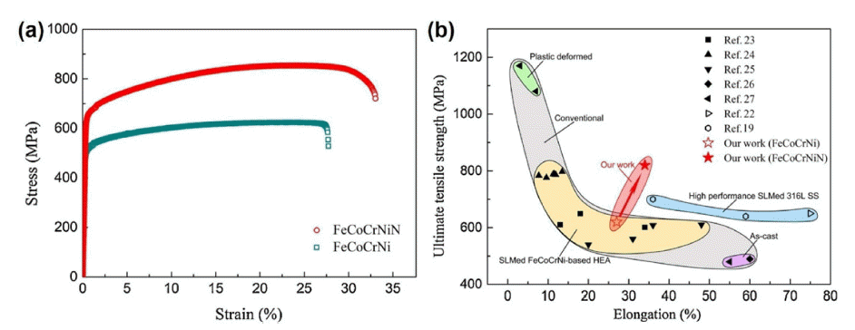
Tensile properties of N-doped and N-free CoCrFeNi HEA fabricated by SLM (reprinted from Ref. [54] with permission from Elsevier): (a) Engineering stress-strain curves for N-free and N-doped HEAs, and (b) Ashby plot of the ultimate tensile strength vs. tensile elongation for SLM-processed singe FCC HEAs, wrought single FCC HEAs, and SLM-processed 316 L stainless steels.
Fig. 15

Microstructural evolution in the SLM-processed 1%C-CoCrFeMnNi HEA (reprinted from Ref. [53] with permission from Elsevier): (a) EBSD IPF map of plane perpendicular to laser scan direction. (b) A schematic of the mechanism of solidification cell growth and texture evolution within a melt pool. (c) EBSD IPF map of the plane perpendicular building direction, and (d) the schematic illustration indicating simplified grain structure in the SLM-processed alloy. Note that the major direction of the laser-scanning is indicated as X direction and the direction parallel to hatch spacing is labeled as Y direction. IPF maps are drawn by crystal orientation plotted // building direction (Z direction).
Fig. 16

Heterogeneous anisotropic grain structure-induced anisotropic plasticity of SLM-processed 1%C-CoCrFeMnNi HEA (reprinted from Ref. [53] with permission from Elsevier): (a) Loading-unloading-reloading (LUR) curves, and (b) back stress contribution to flow stress of the as-built samples in different tensile directions. Note that the laser-scanning direction, the direction parallel to hatch spacing, and the building direction are denoted by X, Y, and Z directions, respectively. (c) EBSD KAM value evolution as a function of local strain of the samples deformed along different tensile axes, and (d, e) EBSD KAM maps of the Z plane of the SLM-processed alloys deformed along (d) X direction and (e) Y direction. The thick double-headed arrows with red color and blue color in the KAM maps represent tensile loading direction of X and Y directions, respectively.
Fig. 17
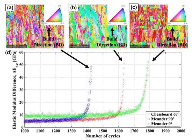
Fatigue properties of SLM-processed CoCrFeMnNi HEA fabricated by different scanning strategies (reprinted from Ref. [55] with permission from Elsevier): EBSD IPF maps of lateral surface of the as-built samples fabricated by (a) the meander 90° scan, (b) the chessboard 67° scan, (c) the meander 0° scan (no rotation). (d) Evolution of the apparent elastic modulus difference for the samples fabricated by the different scanning strategies.
Fig. 18
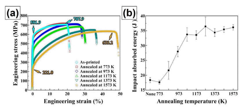
Effect of heat treatment on mechanical properties of SLM-processed CoCrFeMnNi HEA (reprinted from Ref. [63] with permission from Elsevier): (a) Tensile properties and (b) impact toughness of the samples after post heat treatment at different temperatures [63].
Fig. 19
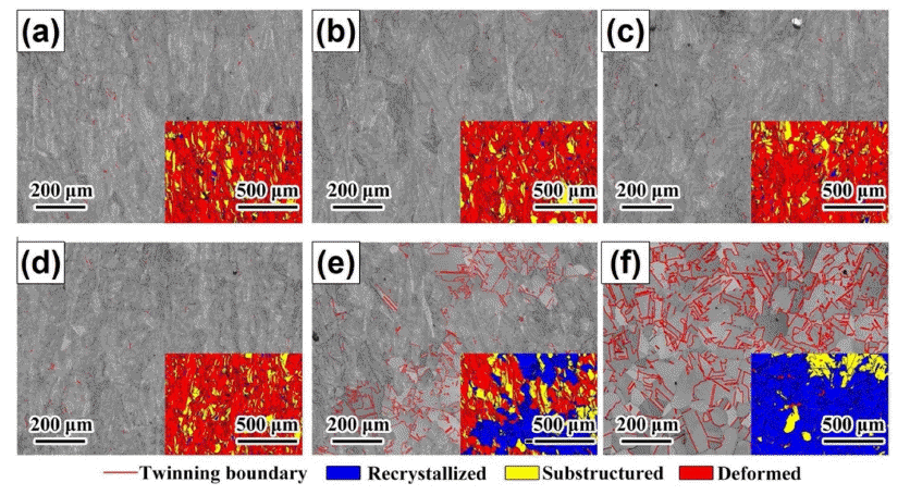
Microstructure of SLM-processed CoCrFeMnNi HEA after post heat treatment at different temperatures (reprinted from Ref. [63] with permission from Elsevier): EBSD IQ maps (insets: recrystallization distribution maps) of the (a) as-built sample and the samples after 2 h of annealing heat treatment at (b) 500°C, (c) 700°C, (d) 900°C, (e) 1100°C, and (f) 1300°C.
Fig. 20
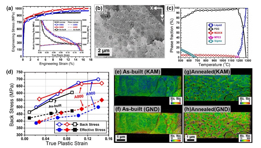
Effect of heat treatment on microstructure and tensile properties of SLM-processed 1%C-CoCrFeMnNi HEA (reprinted from Ref. [64] with permission from Elsevier): (a) Engineering stress-strain curves drawn up to necking of the asbuilt sample and the samples annealed at 800oC (A800) and A900oC (A900). The inset in (a) presents work hardening rate and true stress as a function of true plastic strain of three different samples. (b) SEM BSE micrograph of the annealed sample. (c) Equilibrium phase diagram of the 1%C-CoCrFeMnNi HEA calculated by Thermocalc software. (d) Evolution of back stress and effective stress as a function of true plastic strain of the samples. TKD (e, g) KAM maps and (f, h) GND maps of tensiledeformed microstructures at local strain of 20% in the (e, f) as-built and (g, h) annealed samples. The color bar in the GND maps indicates the range of GND density from 10 × 1012 m-2 to 800 × 1012 m-2.
Fig. 21

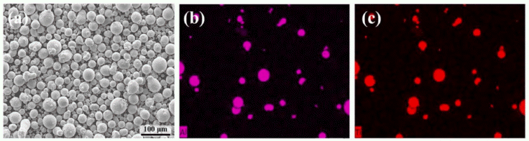
Mixture of CoCrFeMnNi alloy powder and TiAl powder (reprinted from Ref. [65] with permission from Elsevier): (a) SEM micrograph showing the HEA-TiAl mixed powder, and EDS mapping results showing the (b) Ti and (c) Al elements in the powder.
Fig. 22

Various types of precipitates in the SLMed CoCrFeMnNi + TiAl sample after HIP and aging treatment (reprinted from Ref. [65] with permission from Elsevier): (a) HRTEM image taken along [011] zone axis showing the detailed structure in the γ matrix with the FFT pattern in the inset indicating the presence of ordered L12 superlattice structure; (b) Inverse FFT image obtained using {100} and {011} superlattice reflections in (a) showing the presence of long-range ordered L12 domains; Brightfield (BF) TEM micrographs and selected area diffraction patterns (SADP) showing the presence of (c) polygonal α-Al2O3 precipitates in the matrix, (d) σ phase along grain boundaries, (e) B2 precipitates, (f) BCC (Ni,Co)2TiAl Heusler particles and (g) cuboidal γ-TiAl particles in the matrix; (h) HRTEM image and FFT pattern of the γ-TiAl particle shown in (g). The arrow in (g) refers to a superlattice diffraction spot.
Fig. 23

Effect of heat treatment on tensile properties of SLM-processed CoCrFeMnNi-TiAl sample (reprinted from Ref. [65] with permission from Elsevier): (a) Engineering tensile stress-strain curves, (b) true stress-strain curves and strain hardening rate-true strain curves for the as-built sample and the sample after HIP + Ageing treatment.
- 극심한 열이력을 동반하는 SLM 공정은 FCC 단상의 단 조로운 미세조직을 지닌 CoCrFeMnNi 합금에 수 나노 단위 에서 수백 마이크로미터 단위까지의 계층적이며 불균질한 미세조직을 유발한다. 이러한 SLM 조형체의 독특한 미세 조직학적 양상은 단지 연질의 CoCrFeMnNi 합금에 대한 항 복강도 향상뿐만 아니라, 기존 공법재에서 물성 향상을 위 해 제어 가능하였던 제한적인 미세조직학적 변수(결정립도, 전위밀도 등)에서 나노 편석, 나노석출물, 나노 전위셀, 불 균질 결정립 형상 및 배열, 용융풀 등 미세조직학적 제어변 수를 더욱 확장시킬 수 있다는 점에서 큰 의의를 가진다.
- 이러한 SLM 공법 기반의 미세조직 최적화를 통한 맞춤 형 물성 구현을 위해선 각 미세조직학적 인자들이 소재 변형 단계별 미치는 영향과 서로 간의 상호작용에 대한 정량적인 이해가 요구된다. 이때, CoCrFeMnNi 합금은 SLM 공정 적용성이 우수함에 따라 이를 연구하기 위한 건전한 시편 제조가 용이하며, 공정 도중 상변태 및 이차 상 석출 없이 안정한 FCC 단상을 나타낼 수 있음에 따라 그림 24의 개략도와 같이 SLM 조형체의 공정 조건 및 첨 가 원소에 따른 미세조직-기계적물성 상관관계에 대한 기 초 연구들이 활발하게 이루어져 왔다. 이때, 본 리뷰 논문 에서 소개한 CoCrFeMnNi계 합금 기반 SLM 조형체들에 대한 다수의 연구사례들은 SLM 기반 미세조직 제어를 통 한 소재 물성 향상 전략에 대한 가이드라인들을 제공해주 었으며, 이러한 데이터베이스를 기반으로 더욱 소재적인 측면에서의 SLM 공정 연구들이 더욱 활발히 수행될 것으 로 전망할 수 있다. 특히, 지금까지의 물성 한계를 뛰어넘 는 새로운 조성의 신합금이 계속해서 개발되고 있는 고엔 트로피합금 학계에서는 더욱 다양한 조성의 합금들에 대 한 SLM 적용 연구가 수행될 것으로 전망할 수 있다. 즉, 현재 많은 관심을 받고 있는 초고강도의 다상 고엔트로피 합금, 준안정 고엔트로피합금, 초내열/내화 고엔트로피합 금 등에 대해 공정 간소화 및 물성 한계 돌파를 위한 SLM 공법 적용 연구 사례는 계속 증가할 것이며, 향후에 는 SLM 공정에 따른 소재 미세조직 변화 및 특성 변화에 대한 면밀한 이해를 기반으로 적층제조 전용 신조성의 고 엔트로피합금 개발 등의 연구가 수행될 것으로 전망한다.
3. 결 론
-
Acknowledgements
- This work was supported by Principal R&D Project (Grant No. PNK8290) of the Korean Institute of Materials Science (KIMS). This work was also supported by Basic Research Program (Development of Magnetic Powders Specialized for 3D Printing for Next-generation Highefficiency Motors) of Korea Institute of Machinery and Materials (KIMM).
Acknowledgemensts
- 1. D. Raabe, C. C. Tasan and E. A. Olivetti: Nature, 575 (2019) 64..ArticlePubMed
- 2. Y.-J. Liang, L. Wang, Y. Wen, B. Cheng, Q. Wu, T. Cao, Q. Xiao, Y. Xue, G. Sha, Y. Wang, Y. Ren, X. Li, L. Wang, F. Wang and H. Cai: Nat. Commun., 9 (2018) 4063..ArticlePubMedPMC
- 3. D. B. Miracle: Nat. Commun., 10 (2019) 1805..ArticlePubMedPMC
- 4. O. Bouaziz, J. Moon, H. S. Kim and Y. Estrin: Scr. Mater., 191 (2021) 107..Article
- 5. J. M. Park, D. C. Yang, H. Kim, D. G. Kim, S. Lee, H. S. Kim and S. S. Sohn: Mater. Res. Lett., 9 (2021) 315..Article
- 6. P. Sathiyamoorthi and H. S. Kim: Prog. Mater. Sci., 123 (2020) 100709..Article
- 7. Y. H. Jo, S. Jung, W. M. Choi, S. S. Sohn, H. S. Kim, B. J. Lee, N. J. Kim and S. Lee: Nat. Commun., 8 (2017) 15719..ArticlePubMedPMC
- 8. J. Moon, J. M. Park, J. W. Bae, H.-S. Do, B.-J. Lee and H. S. Kim: Acta Mater., 193 (2020) 71..Article
- 9. J. Moon, J. M. Park, J. W. Bae, N. Kang, J. Oh, H. Shin and H. S. Kim: Scr. Mater., 186 (2020) 24..Article
- 10. J. M. Park, J. Moon, J. W. Bae, J. Jung, S. Lee and H. S. Kim: Mater. Sci. Eng. A, 728 (2018) 251..Article
- 11. B. Cantor: Prog. Mater. Sci., 120 (2021) 100754..Article
- 12. G. Laplanche, A. Kostka, O. M. Horst, G. Eggeler and E. P. George: Acta Mater., 118 (2016) 152..Article
- 13. B. Gludovatz, A. Hohenwarter, D. Catoor, E. H. Chang, E. P. George and R. O. Ritchie: Science, 345 (2014) 1153..ArticlePubMed
- 14. H. Thota, R. Jeyaraam, L. R. Bairi, A. S. Tirunilai, A. Kauffmann, J. Freudenberger, M. Heilmaier, S. Mandal and S. S. Vadlamani: J. Alloys. Compd., 888 (2021) 161500..Article
- 15. Y. Zhao, D. H. Lee, M. Y. Seok, J. A. Lee, M. P. Phaniraj, J. Y. Suh, H. Y. Ha, J. Y. Kim, U. Ramamurty and J.-I. Jang: Scr. Mater., 135 (2017) 54..Article
- 16. H. Luo, Z. Li and D. Raabe: Sci. Rep., 7 (2017) 9892. .ArticlePubMedPMC
- 17. J. M. Park, J. Moon, J. W. Bae, M. J. Jang, J. Park, S. Lee and H. S. Kim: Mater. Sci. Eng. A, 719 (2018) 155..Article
- 18. M. J. Yao, K. G. Pradeep, C. C. Tasan and D. Raabe: Scr. Mater., 72-73 (2014) 5..Article
- 19. M. Vaidya, K. G. Pradeep, B. S. Murty, G. Wilde and S. V. Divinski: Acta Mater., 146 (2018) 211..Article
- 20. J. G. Gigax, O. El-Atwani, Q. McCulloch, B. Aytuna, M. Efe, S. Fensin, S. A. Maloy and N. Li: Scr. Mater., 178 (2020) 508..Article
- 21. J. Y. Ko and S. I. Hong: J. Alloys. Compd., 743 (2018) 115..Article
- 22. J. M. Park, J. Moon, J. W. Bae, D. H. Kim, Y. H. Jo, S. Lee and H. S. Kim: Mater. Sci. Eng. A, 746 (2019) 443..Article
- 23. P. Wang, J. Chen, B. Sun, D. Zhu and Y. Cao: Mater. Sci. Eng. A, 840 (2022) 142880..Article
- 24. P. Asghari-Rad, P. Sathiyamoorthi, N. T. Nguyen, A. Zargaran, T. S. Kim and H. S. Kim: Scr. Mater., 190 (2021) 69..Article
- 25. Z. Zhang, X. Zhai, G. Chen, X. Chen and K. Ameyama: Scr. Mater., 213 (2022) 114591..Article
- 26. S. Gorsse, C. Hutchinson, M. Gouné and R. Banerjee: Sci. Technol. Adv. Mater., 18 (2017) 584..ArticlePubMedPMC
- 27. G. M. Karthik and H. S. Kim: Met. Mater. Int., 27 (2021) 1..Article
- 28. A. O. Moghaddam, N. A. Shaburova, M. N. Samodurova, A. Abdollahzadeh and E. A. Trofimov: J. Mater. Sci. Tech., 77 (2021) 131..Article
- 29. M. J. Hwang and J. Cho: J. Welding and Joining, 32 (2014) 15..Article
- 30. D. Herzog, V. Seyda, E. Wycisk and C. Elmmelmann: Acta Mater., 117 (2016) 271..Article
- 31. J. M. Park, J. M. Jeon, J. G. Kim, Y. Seong, S. H. Park and H. S. Kim: J. Powder Mater., 25 (2018) 475..Article
- 32. Y. M. Wang, T. Voisin, J. T. Mckeown, J. Ye, N. P. Calta, Z. Li, Z. Zeng, Y. Zhang, W. Chen, T. T. Roehling, R. T. Ott, M. K. Santala, P. J. Depond, M. J. Matthews, A. V. Hamza and T. Zhu: Nat. Mat., 17 (2018) 63..ArticlePubMed
- 33. L. Liu, Q. Ding, Y. Zhong, J. Zou, J. Wu, Y.-L. Chiu, J. Li, Z. Zhang, Q. Yu and Z. Shen: Mater. Tod., 21 (2018) 354..Article
- 34. A. Piglione, B. Dovgyy, C. Liu, C. M. Gourlay, P. A. Hooper and M. S. Pham: Mater. Lett., 224 (2018) 22..Article
- 35. B. Dovgyy, A. Piglione, P. A. Hooper and M.-S. Pham: Mater. Des., 194 (2020) 108845..Article
- 36. M.-S. Pham, B. Dovgyy, P. A. Hooper, C. M. Gourlay and A. Piglione: Nat. Commun., 11 (2020) 749..ArticlePubMedPMC
- 37. Z. G. Zhu, Q. B. Nguyena, F. L. Nga, X. H. Anb, X. Z. Liao, P. K. Liaw, S. M. L. Nai and J. Wei: Scr. Mater., 154 (2018) 20..Article
- 38. R. Li, P. Niu, T. Yuan, P. Cao, C. Chen and K. Zhou: J. Alloy. Compd., 746 (2018) 125..Article
- 39. D. Kong, C. Dong, S. Wei, X. Ni, L. Zhang, R. Li, L. Wang, C. Man and X. Li: Addit. Manuf., 38 ( 20 21) 101804..Article
- 40. H. Wang, Z. G. Zhu, H. Chen, A. G. Wang, J. Q. Liu, H. W. Liu, R. K. Zheng, S. M. L. Nai, S. Primig, S. S. Babue, S. P. Ringer and X. Z. Liao: Acta Mater., 196 (2020) 609..Article
- 41. Y. K. Kim, S. Yang and K. A. Lee: Sci. Rep., 10 (2020) 8045..ArticlePubMedPMC
- 42. Y. K. Kim, S. Yang and K. A. Lee: Addit. Manuf., 36 (2020) 101543..Article
- 43. Y. K. Kim, M. S. Baek, S. Yang and K. A. Lee: Addit. Manuf., 38 (2021) 101832..Article
- 44. J. M. Park, J. Choe, J. G. Kim, J. W. Bae, J. Moon, S. Yang, K. T. Kim, J.-H. Yu and H. S. Kim: Mater. Res. Lett., 8 (2020) 1..Article
- 45. J. G. Kim, J. M. Park, J. B. Seol, J. Choe, J.-H. Yu, S. Yang and H. S. Kim: Mater. Sci. Eng. A, 773 ( 20 20) 138726..
- 46. Y.-K. Kim, J. H. Yu, H. S. Kim and K.-A. Lee: Compos. B. Eng., 210 (2021) 108638..Article
- 47. H. Chen, T. Lu, Y. Wang, Y. Liu, T. Shi, K. G. Prashanth and K. Kosiba: Mater. Sci. Eng. A, 833 (2022) 142512..Article
- 48. Y. K. Kim, M. C. Jo and K. A. Lee: J. Mater. Sci. Tech., 97 (2022) 10..Article
- 49. P. Chen, C. Yang, S. Li, M. M. Attallah and M. Yan: Mater. Des., 194 (2020) 108966..Article
- 50. S. Thapliyal, P. Agrawal, P. Agrawal, S. S. Nene, R. S. Mishra, B. A. McWilliams and K. C. Cho: Acta Mater., 219 (2021) 117271..Article
- 51. J. M. Park, P. Asghari-Rad, A. Zargaran, J. W. Bae, J. Moon, J. Choe, S. Yang, J.-H. Yu and H. S. Kim: Acta Mater., 221 (2021) 117426..Article
- 52. J. Wang, J. Zou, H. Yang, Z. Liu and S. Ji: Mater. Sci. Eng. A, 843 (2022) 143129..Article
- 53. J. M. Park, J. Choe, H. K. Park, S. Son, J. Jung, T.-S. Kim, J.-H. Yu, J. G. Kim and H. S. Kim: Addit. Manuf., 35 (2020) 101333..Article
- 54. M. Song, R. Zhou, J. Gu, Z. Wang, S. Ni and Y. Liu: Appl. Mater. Tod., 18 (2020) 100498..Article
- 55. M. Jin, A. Piglione, B. Dovgyy, E. Hosseini, P. A. Hooper, S. R. Holdsworth and M.-S. Pham: Addit. Manuf., 36 (2020) 101584..Article
- 56. M. Zheng, C. Li, L. Zhang, X. Zhang, Z. Ye, X. Yang and J. Gu: Mater. Sci. Eng. A, 840 (2022) 142933. .Article
- 57. S. A. H. Motaman, F. Roters and C. Haase: Acta Mater., 185 (2020) 340..Article
- 58. A. M. Beese, Z. Wang, A. D. Stoica and D. Ma: Nat. Commun., 9 (2018) 2083..ArticlePubMedPMC
- 59. W. Chen, T. Voisin, Y. Zhang, J.-B. Forien, C. M. Spadaccini, D. L. McDowell, T. Zhu and Y. M. Wang: Nat. Commun., 10 (2019) 4338..ArticlePubMedPMC
- 60. C. Wang, X. P. Tan, S. B. Tor and C. S. Lim: Addit. Manuf., 36 (2020) 101538..Article
- 61. J. Jung, J. Na, H. K. Park, J. M. Park, G. Kim, S. Lee and H. S. Kim: npj Comput. Mater., 7 (2021) 1..Article
- 62. J. Qin, F. Hu, Y. Liu, P. Witherell, C. C. L. Wang, D. W. Rosen, T. W. Simpson, Y. Lu and Q. Tang: Addit. Manuf., 52 (2022) 102691..Article
- 63. D. Lin, L. Xu, H. Jing, Y. Han, L. Zhao and F. Minami: Addit. Manuf., 32 (2020) 101058..Article
- 64. J. M. Park, E. S. Kim, H. Kwon, P. Sathiyamoorthi, K. T. Kim, J. H. Yu and H. S. Kim: Addit. Manuf., 47 (2021) 102283..Article
- 65. X. Wang, X. Pan, P. Sun and C. Qiu: Mater. Sci. Eng. A, 831 (2022) 142285..Article
Figure & Data
References
Citations
Citations to this article as recorded by 
- Thermodynamic and Electronic Descriptor-Driven Machine Learning for Phase Prediction in High-Entropy Alloys: Experimental Validation
Nguyen Lam Khoa, Nguyen Duy Khanh, Hoang Thi Ngoc Quyen, Nguyen Thi Hoang, Oanh, Le Hong Thang, Nguyen Hoa Khiem, Nguyen Hoang Viet
Journal of Powder Materials.2025; 32(3): 191. CrossRef - Investigation of effects of process parameters on microstructure and fracture toughness of SLM CoCrFeMnNi
Joseph Agyapong, Diego Mateos, Aleksander Czekanski, Solomon Boakye-Yiadom
Journal of Alloys and Compounds.2024; 987: 173998. CrossRef - Cryogenic Tensile Behavior of Ferrous Medium-entropy Alloy Additively Manufactured by Laser Powder Bed Fusion
Seungyeon Lee, Kyung Tae Kim, Ji-Hun Yu, Hyoung Seop Kim, Jae Wung Bae, Jeong Min Park
journal of Korean Powder Metallurgy Institute.2024; 31(1): 8. CrossRef - Data-driven Approach to Explore the Contribution of Process Parameters for Laser Powder Bed Fusion of a Ti-6Al-4V Alloy
Jeong Min Park, Jaimyun Jung, Seungyeon Lee, Haeum Park, Yeon Woo Kim, Ji-Hun Yu
journal of Korean Powder Metallurgy Institute.2024; 31(2): 137. CrossRef - Cryogenic tensile behavior of carbon-doped CoCrFeMnNi high-entropy alloys additively manufactured by laser powder bed fusion
Haeum Park, Hyeonseok Kwon, Kyung Tae Kim, Ji-Hun Yu, Jungho Choe, Hyokyung Sung, Hyoung Seop Kim, Jung Gi Kim, Jeong Min Park
Additive Manufacturing.2024; 86: 104223. CrossRef - Microstructural evolution and high strain rate deformation response of SLM-printed CoCrFeMnNi after annealing and deep-cryogenic treatment
Joseph Agyapong, Aleksander Czekanski, Solomon Boakye Yiadom
Materials Characterization.2024; 218: 114506. CrossRef - High-speed manufacturing-driven strength-ductility improvement of H13 tool steel fabricated by selective laser melting
Yeon Woo Kim, Haeum Park, Young Seong Eom, Dong Gill Ahn, Kyung Tae Kim, Ji-hun Yu, Yoon Suk Choi, Jeong Min Park
Powder Metallurgy.2023; 66(5): 582. CrossRef
 ePub Link
-
 Cite this Article
- Cite this Article
-
- Close
- Download Citation
- Close
- Figure
-


- Related articles
-
- Microstruture and Mechanical Properties of Ti.Grade12-Ti/TiN/WC Composite Produced by Spark Plasma Sintering Process
- Microstructure and Mechanical Properties of AA3003 Tube for Heat Exchanger Processed by Floating Plug Drawing
- Effect of Hatch Spacing on the Microstructure and Mechanical Properties of SA508 Gr.3 Steel Fabricated by Laser Powder Bed Fusion
- Microstructure and Mechanical Properties of Laser Powder Bed Fusion 3D-Printed Cu-10Sn Alloy
- Comparative Review of the Microstructural and Mechanical Properties of Ti-6Al-4V Fabricated via Wrought and Powder Metallurgy Processes
Microstructure and Mechanical Properties of CoCrFeMnNi-type High-entropy Alloy Fabricated by Selective Laser Melting: A Review


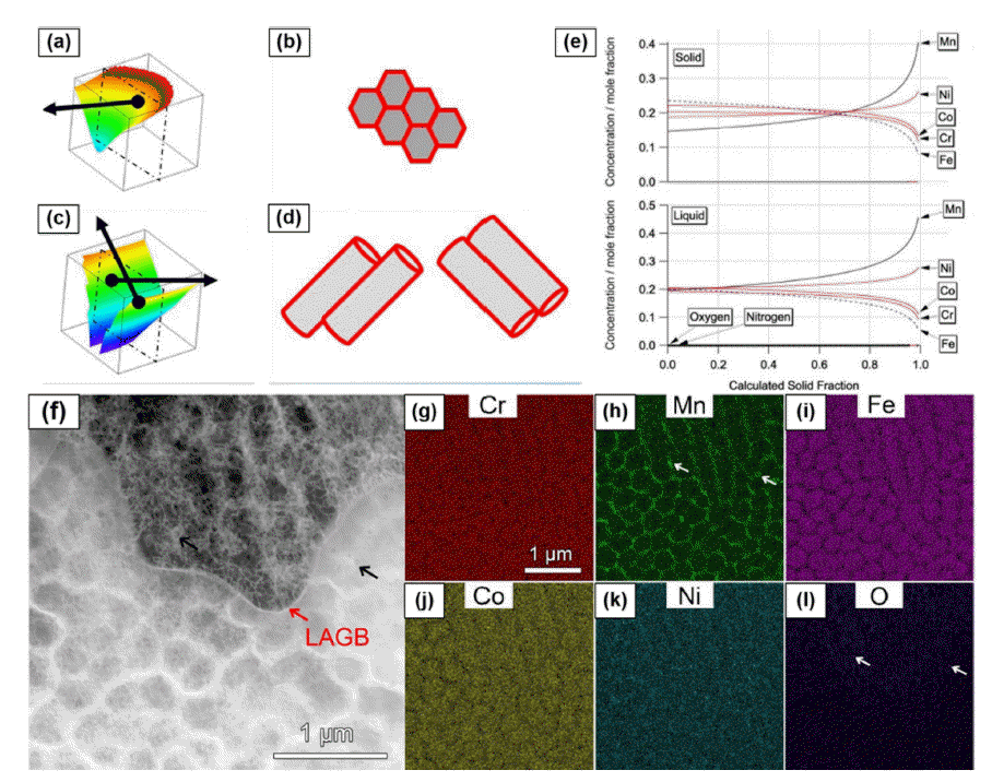

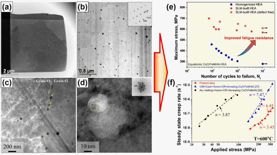

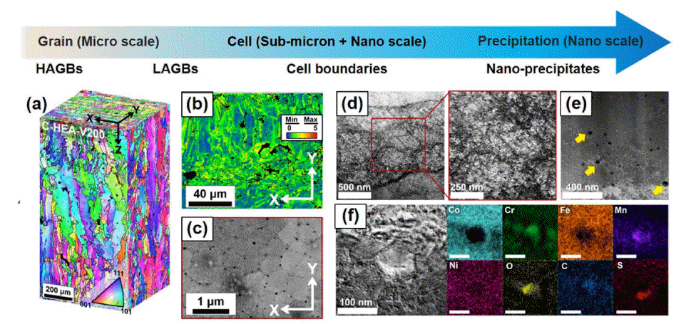
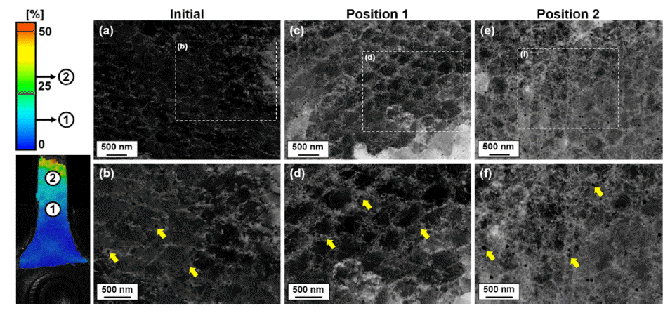

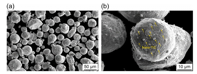
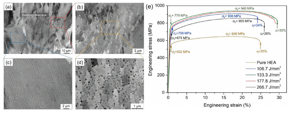


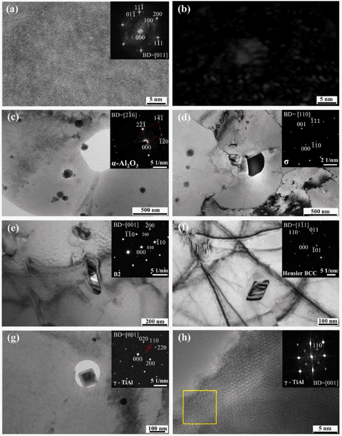
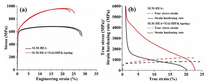

Fig. 1
(a) A schematic of SLM process. (b) SLM-produced brackets (left) on a build plate and finished part (right) for Airbus A350 with topology optimized design resulting in ~30% weight reduction of conventional part (reprinted from [30] with permission from Elsevier). (c) Schematic illustrations indicating the process of tool path generation from CAD data and layer processing for additive manufacturing [29].
Fig. 2
Microstructure of SLM-processed CoCrFeMnNi alloy [34-36] (reproduced with permission from Springer Nature and Elsevier): (a) EBSD-IPF map indicating solidification microstructure of a CoCrFeMnNi HEA processed by SLM in a singletrack building [34]. Note that the blue box marked in (a) was magnified as the SEM micrograph indicating solidification cells at the right side of the SEM micrograph, and a red (<001> orientation) and green (<101> orientation) colored cubic pointed by red arrow represents the crystallographic orientation of a solidification cell indicated in IPF map and SEM micrograph [35]. (b) Variation of microstructure of SLM-processed HEA from the substrate (316L stainless steel) towards the top of the sample [35]. (c) SEM image indicating the side-branching of the solidified grains between adjacent melt tracks [35]. (d) Microstructure of building plane of the samples processed by SLM in different scanning strategies [36]. (e) 3D pseudo microstructure of the CoCrFeMnNi alloy processed by 67° rotation in every layer under chessboard laser scanning patten during SLM [36].
Fig. 3
Hierarchical heterogeneous microstructure of SLM-processed CoCrFeMnNi alloy (reprinted from [37] with permission from Elsevier): (a) OM and (b) SEM images of the lateral surface of the as-built sample. (c) SEM micrograph indicating cell structure. (d) EBSD IPF map, (e) EBSD IQ map indicating high angle grain boundaries (blue lines) and low angle grain boundaries (red lines), and (f) EBSD KAM amp image of the lateral surface of the as-built sample. (g) Bright-field STEM micrograph showing cell structure, and (h) EDS mapping results for blue box indicated in (g).
Fig. 4
Solidification cell structure of SLM-processed CoCrFeMnNi alloy (reprinted from [40] with permission from Elsevier): (a) Extracted melt pool shape near the rear of the weld pool and direction of the [100]FCC primary solidification direction is expected to be mostly along to the scan direction of SLM. (b) The schematic of solidified cell structure showing honeycomb structure observed in the plane perpendicular to scan direction. (c) Extracted melt pool boundaries along the sides of the melt pools and (d) will appear like dendritic cell in the plane perpendicular to scan direction. (e) Simulated concentrations of alloying elements in liquid and solid as a function of mole fraction solid. (f) A STEM HAADF micrograph image showing cell structure with (g-l) EDS maps of the constituent elements. The two white arrows in the Mn and O maps point to two participates (MnO).
Fig. 5
(a) Tensile properties of SLM-processed CoCrFeMnNi alloy and post heat-treated samples (reprinted from [37] with permission from Elsevier). Deformed microstructure of the alloy produced by SLM [37]: (b) EBSD IPF, (c) EBSD KAM, and (d, e) bright-field STEM micrographs of the tensile-fractured sample.
Fig. 6
Effect of in-situ formed nano-oxide in SLM-processed CoCrFeMnNi alloy (reprinted from Refs. [42] and [43] with permission from Elsevier): (a) TEM, (b) STEM, (c) STEM-HAADF, and (d) HR-TEM micrographs showing nano-sized MnO at cell boundaries in the sample. (e) S-N curves and (f) stress dependencies of steady-state creep rate of SLM-processed CoCrFeMnNi HEAs compared with the wrought alloys.
Fig. 7
Mechanical properties of SLM-processed 1%C-CoCrFeMnNi alloy (reprinted from Ref. [44] with permission from Taylor & Francis): (a) Engineering stress-strain curves of carbon-free (labeled by HEA) and carbon-doped CoCrFeMnNi HEAs (labeled by C-HEA) fabricated by SLM compared with that of the as-cast C-HEA. Note that the laser scanning speeds of 200 mm/s and 600 mm/s used for sample building were indicated by V200 and V600 in the sample name. (b) Ashby plot of the yield strength and tensile elongation for the SLM-processed C-HEA compared with other SLM-processed CoCrFeMnNi-type HEAs.
Fig. 8
Hierarchical heterogeneous microstructure of carbon-doped CoCrFeMnNi alloy fabricated by SLM (reproduced from Ref. [44] with permission from Taylor & Francis): (a) 3D EBSD IPF maps, (b) EBSD KAM map, (c) SEM BSE micrograph, STEM micrograph showing (d) cell structure with (e) nano-precipitates, and (f) EELS mapping results for the nano-precipitate at cell boundaries in the SLM-processed alloy.
Fig. 9
Deformed microstructure of the carbon-doped CoCrFeMnNi HEA processed by SLM (reprinted from Ref. [45] with permission from Elsevier): ECCI results for (a, b) as-built sample, and the tensile deformed samples at (c, d) Position 1 (local strain of 10%) and (e, f): Position 2 (local strain of 25%). The strain distribution from DIC analysis provides local strain of the tensile deformed sample. The yellow arrows in ECCI micrographs indicate the presence of nano-precipitates (dark contrast) at the cell boundaries with dislocations (white contrast).
Fig. 10
Schematic illustrations of (a) temperature vs. exposure time for Cr23C6 formation (sensitization) and (b) the temperature variation as a function of time in a melt pool of exposed by laser beam during SLM in different scanning speed conditions for printing the carbon-doped CoCrFeMnNi HEA.
Fig. 11
(a) SEM micrographs showing the CoCrFeMnNi HEA powder mixed with nano-TiC particle, and the magnified SEM micrograph of the particle (reprinted from Ref. [47] with permission from Elsevier).
Fig. 12
Nano-TiC reinforced CoCrFeMnNi HEA produced by SLM (reprinted from Ref. [47] with permission from Elsevier): (a) SEM image showing cell structure with different morphology, and showing typical morphology of (b) cell and (c) dendritic columnar structure taken from the locations marked with the red and blue rectangle in (a). (d) High magnification SEM image showing nano-precipitates in the composite sample. (e) Tensile properties of TiC-doped and TiC-free HEA produced by SLM in different process parameters.
Fig. 13
Microstructure of N-doped and N-free CoCrFeNi HEA fabricated by SLM (reprinted from Ref. [54] with permission from Elsevier): (a, b) EBSD IPF maps and (c, d) grain size distribution of building plane of (a, c) N-free and (b, d) N-doped HEAs.
Fig. 14
Tensile properties of N-doped and N-free CoCrFeNi HEA fabricated by SLM (reprinted from Ref. [54] with permission from Elsevier): (a) Engineering stress-strain curves for N-free and N-doped HEAs, and (b) Ashby plot of the ultimate tensile strength vs. tensile elongation for SLM-processed singe FCC HEAs, wrought single FCC HEAs, and SLM-processed 316 L stainless steels.
Fig. 15
Microstructural evolution in the SLM-processed 1%C-CoCrFeMnNi HEA (reprinted from Ref. [53] with permission from Elsevier): (a) EBSD IPF map of plane perpendicular to laser scan direction. (b) A schematic of the mechanism of solidification cell growth and texture evolution within a melt pool. (c) EBSD IPF map of the plane perpendicular building direction, and (d) the schematic illustration indicating simplified grain structure in the SLM-processed alloy. Note that the major direction of the laser-scanning is indicated as X direction and the direction parallel to hatch spacing is labeled as Y direction. IPF maps are drawn by crystal orientation plotted // building direction (Z direction).
Fig. 16
Heterogeneous anisotropic grain structure-induced anisotropic plasticity of SLM-processed 1%C-CoCrFeMnNi HEA (reprinted from Ref. [53] with permission from Elsevier): (a) Loading-unloading-reloading (LUR) curves, and (b) back stress contribution to flow stress of the as-built samples in different tensile directions. Note that the laser-scanning direction, the direction parallel to hatch spacing, and the building direction are denoted by X, Y, and Z directions, respectively. (c) EBSD KAM value evolution as a function of local strain of the samples deformed along different tensile axes, and (d, e) EBSD KAM maps of the Z plane of the SLM-processed alloys deformed along (d) X direction and (e) Y direction. The thick double-headed arrows with red color and blue color in the KAM maps represent tensile loading direction of X and Y directions, respectively.
Fig. 17
Fatigue properties of SLM-processed CoCrFeMnNi HEA fabricated by different scanning strategies (reprinted from Ref. [55] with permission from Elsevier): EBSD IPF maps of lateral surface of the as-built samples fabricated by (a) the meander 90° scan, (b) the chessboard 67° scan, (c) the meander 0° scan (no rotation). (d) Evolution of the apparent elastic modulus difference for the samples fabricated by the different scanning strategies.
Fig. 18
Effect of heat treatment on mechanical properties of SLM-processed CoCrFeMnNi HEA (reprinted from Ref. [63] with permission from Elsevier): (a) Tensile properties and (b) impact toughness of the samples after post heat treatment at different temperatures [63].
Fig. 19
Microstructure of SLM-processed CoCrFeMnNi HEA after post heat treatment at different temperatures (reprinted from Ref. [63] with permission from Elsevier): EBSD IQ maps (insets: recrystallization distribution maps) of the (a) as-built sample and the samples after 2 h of annealing heat treatment at (b) 500°C, (c) 700°C, (d) 900°C, (e) 1100°C, and (f) 1300°C.
Fig. 20
Effect of heat treatment on microstructure and tensile properties of SLM-processed 1%C-CoCrFeMnNi HEA (reprinted from Ref. [64] with permission from Elsevier): (a) Engineering stress-strain curves drawn up to necking of the asbuilt sample and the samples annealed at 800oC (A800) and A900oC (A900). The inset in (a) presents work hardening rate and true stress as a function of true plastic strain of three different samples. (b) SEM BSE micrograph of the annealed sample. (c) Equilibrium phase diagram of the 1%C-CoCrFeMnNi HEA calculated by Thermocalc software. (d) Evolution of back stress and effective stress as a function of true plastic strain of the samples. TKD (e, g) KAM maps and (f, h) GND maps of tensiledeformed microstructures at local strain of 20% in the (e, f) as-built and (g, h) annealed samples. The color bar in the GND maps indicates the range of GND density from 10 × 1012 m-2 to 800 × 1012 m-2.
Fig. 21
Mixture of CoCrFeMnNi alloy powder and TiAl powder (reprinted from Ref. [65] with permission from Elsevier): (a) SEM micrograph showing the HEA-TiAl mixed powder, and EDS mapping results showing the (b) Ti and (c) Al elements in the powder.
Fig. 22
Various types of precipitates in the SLMed CoCrFeMnNi + TiAl sample after HIP and aging treatment (reprinted from Ref. [65] with permission from Elsevier): (a) HRTEM image taken along [011] zone axis showing the detailed structure in the γ matrix with the FFT pattern in the inset indicating the presence of ordered L12 superlattice structure; (b) Inverse FFT image obtained using {100} and {011} superlattice reflections in (a) showing the presence of long-range ordered L12 domains; Brightfield (BF) TEM micrographs and selected area diffraction patterns (SADP) showing the presence of (c) polygonal α-Al2O3 precipitates in the matrix, (d) σ phase along grain boundaries, (e) B2 precipitates, (f) BCC (Ni,Co)2TiAl Heusler particles and (g) cuboidal γ-TiAl particles in the matrix; (h) HRTEM image and FFT pattern of the γ-TiAl particle shown in (g). The arrow in (g) refers to a superlattice diffraction spot.
Fig. 23
Effect of heat treatment on tensile properties of SLM-processed CoCrFeMnNi-TiAl sample (reprinted from Ref. [65] with permission from Elsevier): (a) Engineering tensile stress-strain curves, (b) true stress-strain curves and strain hardening rate-true strain curves for the as-built sample and the sample after HIP + Ageing treatment.
Fig. 24
A schematic diagram for the research strategy to establish the relationship of microstructure-mechanical properties of CoCrFeMnNi-type HEA produced by SLM (reproduced from Ref. [44], [53], and [64] with permission from Taylor & Francis and Elsevier).
Fig. 1
Fig. 2
Fig. 3
Fig. 4
Fig. 5
Fig. 6
Fig. 7
Fig. 8
Fig. 9
Fig. 10
Fig. 11
Fig. 12
Fig. 13
Fig. 14
Fig. 15
Fig. 16
Fig. 17
Fig. 18
Fig. 19
Fig. 20
Fig. 21
Fig. 22
Fig. 23
Fig. 24
Microstructure and Mechanical Properties of CoCrFeMnNi-type High-entropy Alloy Fabricated by Selective Laser Melting: A Review
TOP
