 KPMI


Articles
- Page Path
- HOME > J Powder Mater > Volume 33(5); 2026 > Article
-
Research Article
- Role of Oxides in the Tensile Properties of Pure Copper Processed by Laser Powder Bed Fusion
- Haeum Park1,2,†, Ki Jong Kim3,4,†, Yeon Woo Kim1, Hahun Lee5, Kichul Park3, Yerim Kim3, Kee-Ahn Lee4, Seok Su Sohn2,5, Jeong Min Park1,2,6,7,8,*
-
Journal of Powder Materials 2026;33(5):372-378.
DOI: https://doi.org/10.4150/jpm.2026.00248
Published online: October 31, 2026
1Division of Nano Materials, Korea Institute of Materials Science (KIMS), Changwon 51508, Republic of Korea
2Department of Advanced Future Convergence Materials, Korea University, Seoul 02841, Republic of Korea
3Metal 3D Printing Team, Partners Lab Co., Ltd., Seoul 02841, Republic of Korea
4Department of Materials Science and Engineering, Inha University, Incheon 22212, Republic of Korea
5Department of Materials Science and Engineering, Korea University, Seoul 02841, Republic of Korea
6Center for 3D Printing Process, Korea Institute of Materials Science (KIMS), Changwon 51508, Republic of Korea
7Center for Advanced Functional Powder Materials, Korea Institute of Materials Science (KIMS), Changwon 51508, Republic of Korea
8Division of AI for Materials Research, Korea Institute of Materials Science (KIMS), Changwon 51508, Republic of Korea
- *Corresponding Jeong Min Park Tel: +82-55-280-3502; E-mail: jmpark@kims.re.kr
- †These authors contributed equally to this work.
© The Korean Powder Metallurgy & Materials Institute
This is an Open Access article distributed under the terms of the Creative Commons Attribution Non-Commercial License (http://creativecommons.org/licenses/by-nc/4.0/) which permits unrestricted non-commercial use, distribution, and reproduction in any medium, provided the original work is properly cited.
- 158 Views
- 3 Download

Abstract
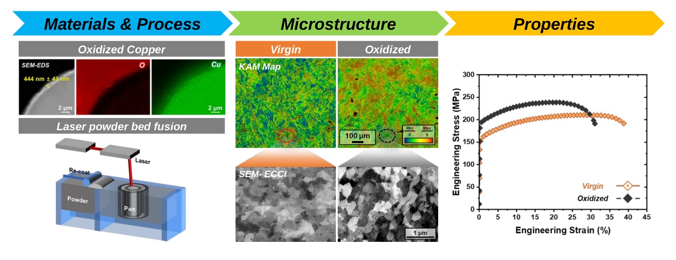
- Oxide particles can affect the tensile properties of pure Cu processed by laser powder bed fusion (LPBF) through their interactions with dislocations. This study compared the microstructure and tensile behavior of samples produced from virgin and oxidized pure Cu powders under identical LPBF conditions. The two samples had comparable relative densities above 98.7% and similar grain sizes. Yield and ultimate tensile strengths were nevertheless higher in the oxidized sample, whereas uniform and total elongations were lower. Backscattered-electron imaging and image analysis indicated that oxide particles occupied a higher apparent area fraction in the examined fields of the oxidized sample; electron backscatter diffraction analysis also showed higher kernel average misorientation. Together, these observations suggest two possible roles for oxide particles: acting as dispersed obstacles to dislocation motion and promoting dislocation retention during LPBF, consistent with the higher kernel average misorientation. The strength increase may therefore reflect both particle strengthening and a greater contribution from dislocation strengthening.
- The expansion of electrified transportation and energy systems is increasing the demand for compact electric motors and power electronic devices with high power density [1, 2]. These systems include motor windings and busbars for current transport and heat exchangers and cooling structures for heat removal [1-3]. Pure copper is widely used in these components because of its excellent electrical and thermal conductivities [1-4].
- The efficiency of these components also depends on their geometry [1, 2]. Optimized conductor paths and internal cooling channels can improve current delivery and heat removal but are difficult to fabricate using conventional processes [1-3, 5, 6]. Laser powder bed fusion (LPBF) enables the direct fabrication of complex copper components with internal features [4-6].
- Realizing these geometrical advantages in pure copper requires reliable LPBF processing. However, its low absorptivity for near-infrared fiber lasers and high thermal conductivity limit energy coupling, resulting in unstable melting and lack-of-fusion defects [4, 6-10]. Previous studies have addressed this problem using higher laser power, green or blue laser sources, and optimized processing parameters and scan strategies [6-14]. On the other hand, feedstock modification provides another route for improving laser-energy absorption [15-20]. Jadhav et al. [17] reported that surface oxidation increased the optical absorption of copper powder and enabled the fabrication of dense copper using a near-infrared laser. However, virgin and oxidized powders were not compared side-by-side under identical LPBF conditions.
- In this study, both powders were processed under identical LPBF conditions, and the relative density, tensile behavior, grain structure, local misorientation, and oxide particle distribution of the fabricated samples were comparatively evaluated.
1. Introduction
- 2.1. Sample fabrication
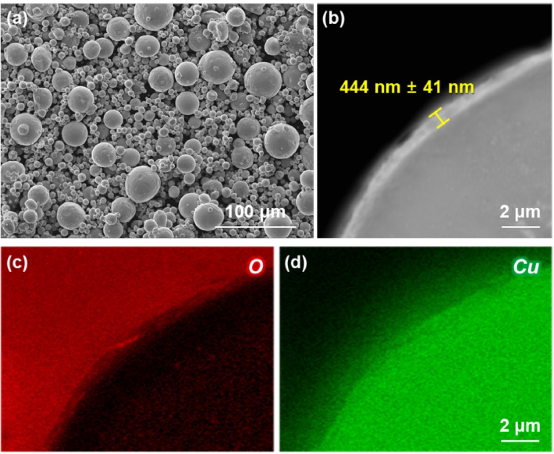
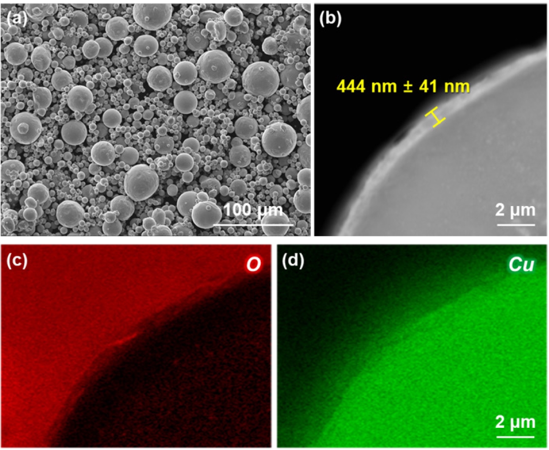
- Spherical pure Cu powder (CuCP; EOS GmbH, Germany) with particle sizes of 10–55 μm was used as the starting material. Two powder conditions were considered: virgin powder and powder with a surface oxide layer, hereafter referred to as the virgin and oxidized powders, respectively. The morphology of the oxidized powder was observed using field-emission scanning electron microscopy (FE-SEM; JSM-7001F, JEOL, Japan). The surface oxide layer was examined by cross-sectional backscattered-electron (BSE) imaging, and the distributions of O and Cu near the powder surface were analyzed using energy-dispersive X-ray spectroscopy (EDS), as shown in Fig. 1.
- Cuboidal samples (12 × 12 × 12 mm³) were fabricated separately from the virgin and oxidized powders using an EOS M290 LPBF system (EOS GmbH, Germany). A laser power of 900 W and a scan speed of 700 mm s⁻¹ were applied to both powder conditions. The as-printed samples fabricated from the virgin and oxidized powders are denoted Virgin and Oxidized, respectively.
- 2.2. Microstructural characterization
- The surface morphologies of the virgin and oxidized powders were examined using scanning electron microscopy (SEM). For relative-density measurements, the fabricated samples were mechanically polished, and optical microscopy images were obtained from their cross-sections. The relative density was calculated as the ratio of the solid area to the total analyzed area using image analysis.
- The crystallographic orientation and grain structure of the fabricated samples were characterized using electron backscatter diffraction (EBSD). The EBSD maps were acquired with a step size of 1.5 μm. Individual grains were reconstructed from the orientation maps, and the grain size was evaluated using the equivalent circle diameter (ECD). Kernel average misorientation (KAM) maps were also used to compare the local lattice misorientation of the samples. The size and distribution of oxide particles were further examined using backscattered-electron (BSE) imaging.
- 2.3. Mechanical testing
- For tensile testing, flat miniature tensile specimens with an overall length of 10 mm and a gage length of 3 mm were machined from the LPBF-processed samples. Miniature tensile specimens with a 3 mm gage length have previously been used to evaluate the mechanical properties of metallic materials [21, 22]. The tensile axis was oriented perpendicular to the build direction (BD). Uniaxial tensile tests were conducted at a strain rate of 1 × 10⁻³ s⁻¹ and 298 K using an Instron 3367 testing machine equipped with a digital image correlation system (DIC; ARAMIS 2D, GOM, Germany) [23]. The yield strength was determined using the 0.2% offset method described in ASTM E8/E8M [24]. One tensile specimen was tested for each condition owing to limited specimen availability.
2. Experimental procedure
- 3.1. Relative density
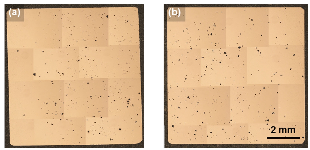
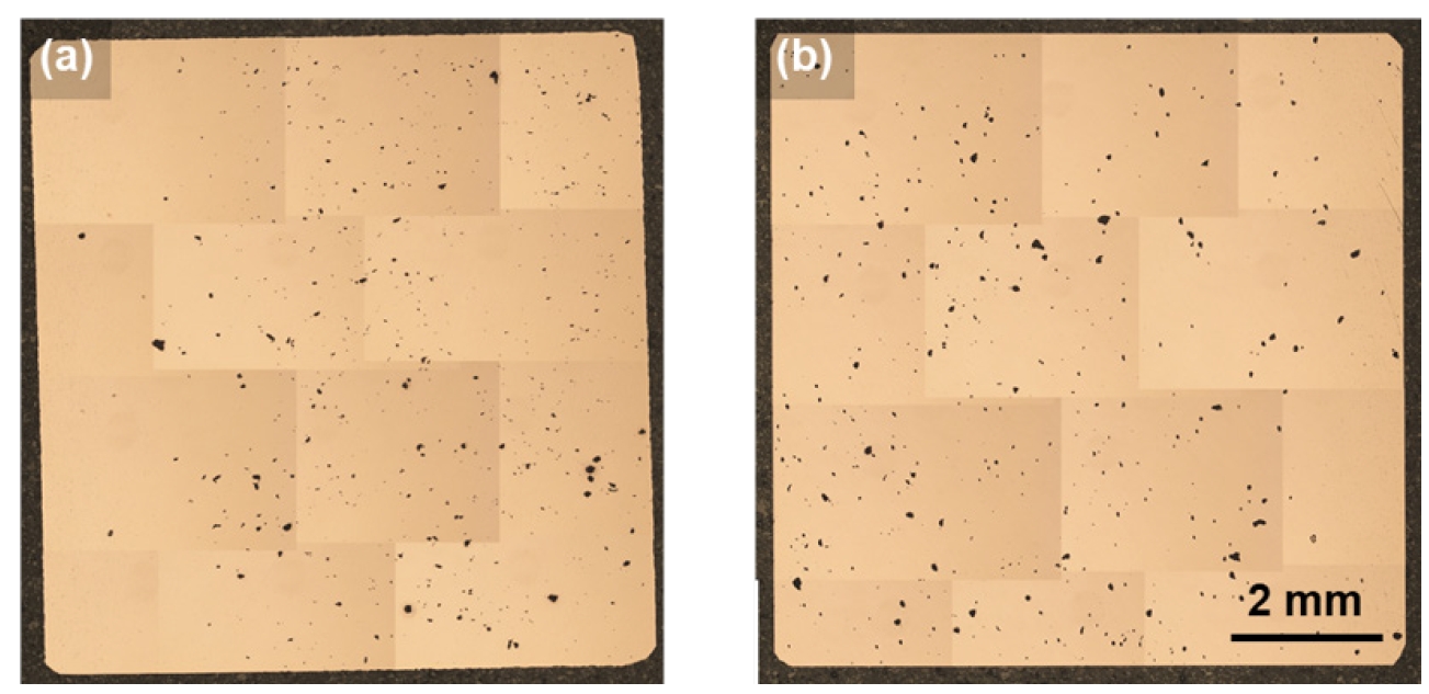
- Figure 2 shows cross-sectional optical micrographs of the (a) Virgin and (b) Oxidized samples. Isolated pores were observed in both samples. The relative densities of the Virgin and Oxidized samples were 98.8% and 98.7%, respectively. These results indicate comparable densification under the applied LPBF condition.
- 3.2. Tensile behavior
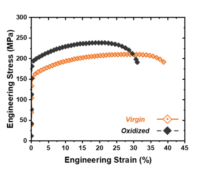
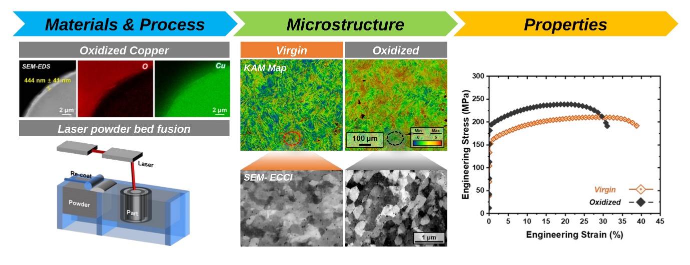
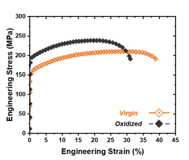
- Despite their comparable relative densities, the Oxidized sample exhibited higher tensile strength than the Virgin sample. Figure 3 presents the engineering stress–strain curves, and the tensile properties are summarized in Table 1. The yield strength (YS) increased from 157 MPa in the Virgin sample to 195 MPa in the Oxidized sample, corresponding to an increase of approximately 24.2%. The ultimate tensile strength (UTS) also increased from 211 to 239 MPa, representing an increase of approximately 13.3%. In contrast, the total elongation (T. El.) decreased from 38.8% to 28.6%, while the uniform elongation (U. El.) decreased from 27.4% to 19.6%.
- Fine oxide particles dispersed in a Cu matrix can impede dislocation motion and increase the strength of LPBF-fabricated Cu [17, 20]. In addition to their strengthening effect, oxide particles may facilitate microvoid nucleation through local stress concentrations at particle–matrix interfaces during tensile deformation. Such damage could contribute to the lower total elongation of the Oxidized sample, although particle-associated void formation was not directly confirmed in this study. The microstructural origin of this difference was further examined using EBSD and BSE imaging.
- 3.3. Microstructural characteristics
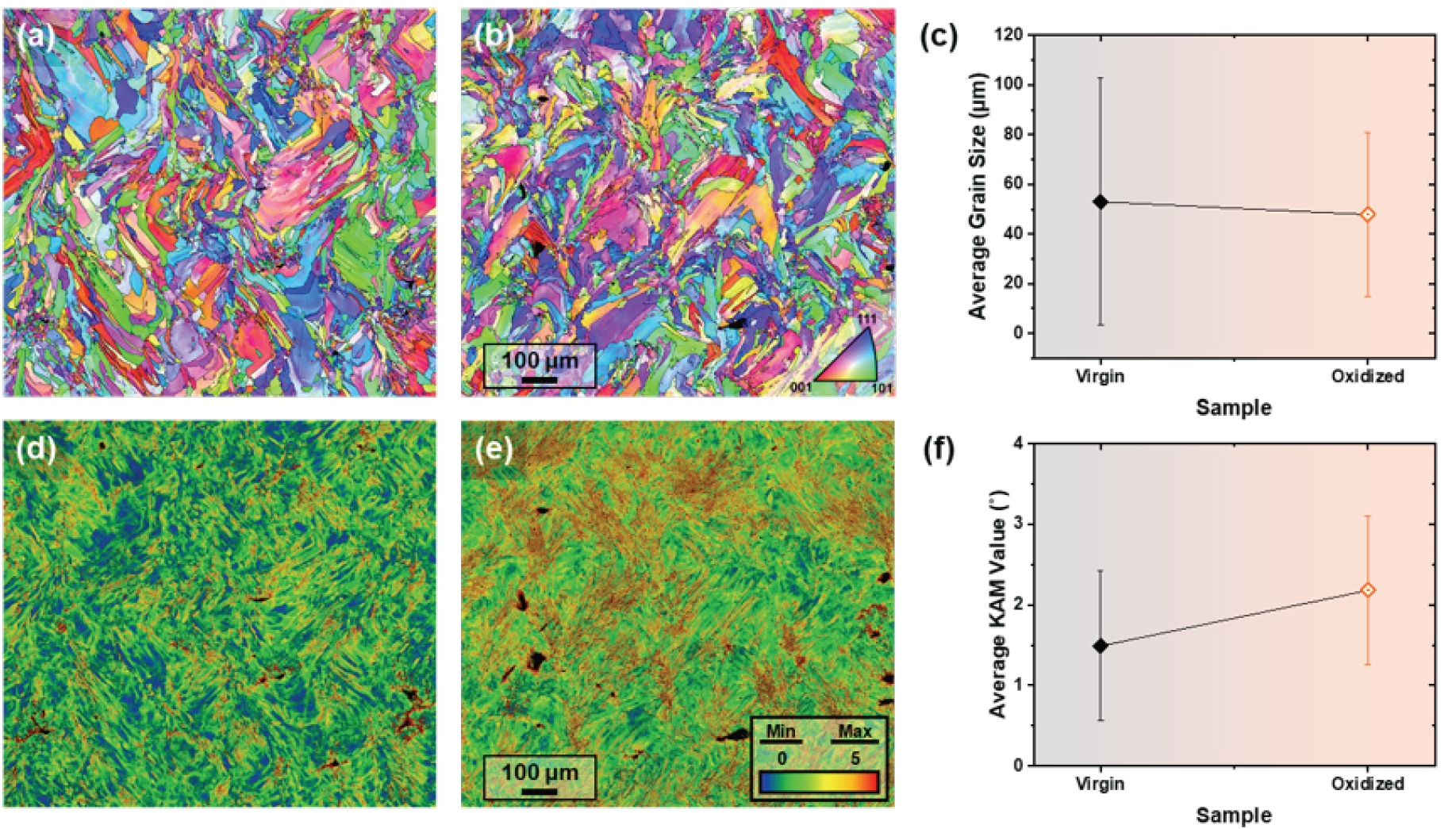
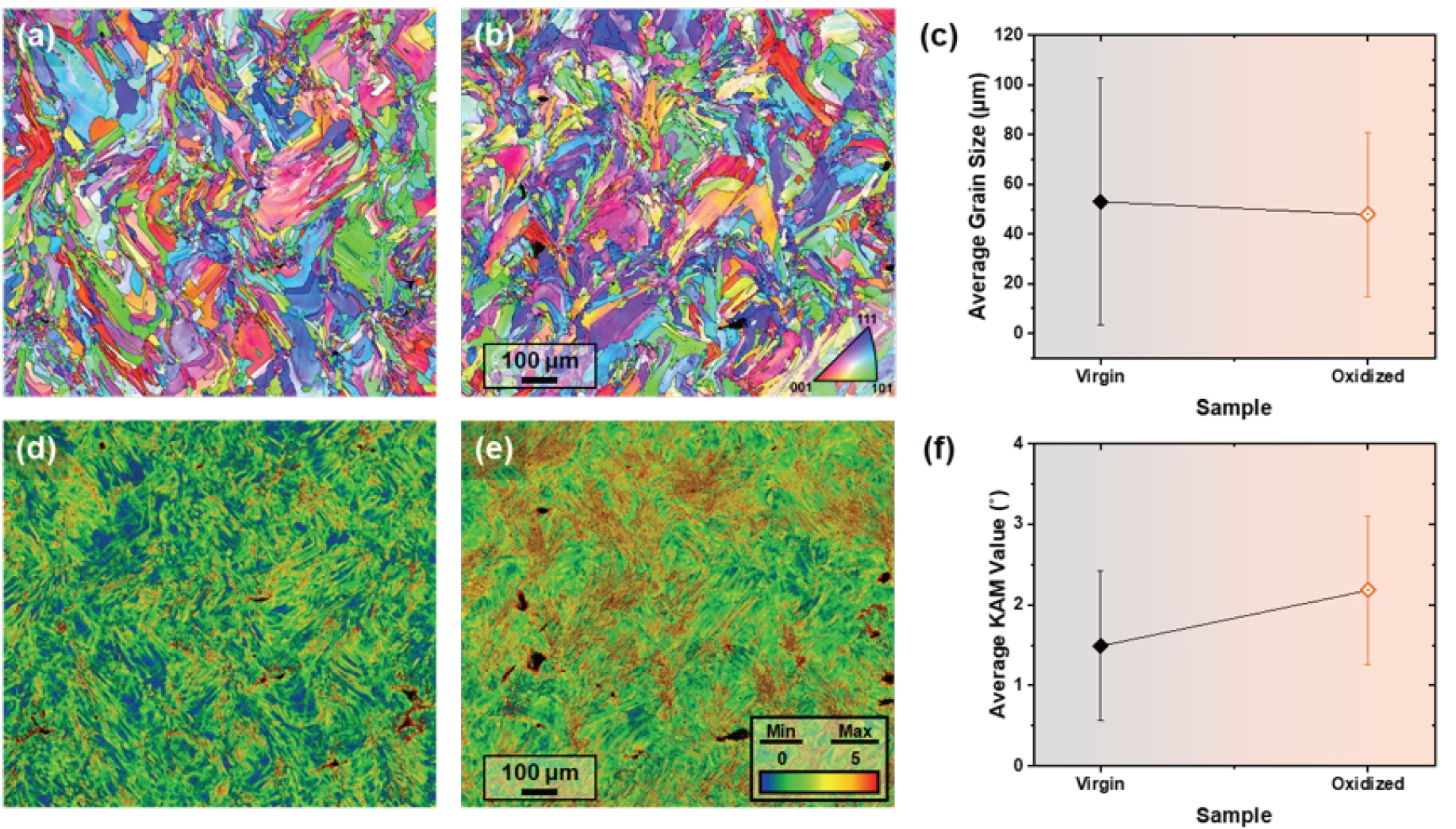
- Figure 4 shows the EBSD inverse pole figure (IPF) and KAM maps of the Virgin and Oxidized samples. The mean grain sizes, determined using the equivalent circle diameter, were 53 ± 50 μm and 48 ± 33 μm for the Virgin and Oxidized samples, respectively. Although the Oxidized sample had a slightly smaller mean grain size, both samples showed broad grain-size distributions. The limited difference in grain size is unlikely to fully account for the increase in YS.
- The YS of a metallic material can be expressed as the sum of the matrix friction stress and the contributions from solid-solution, grain-boundary, dislocation, and particle strengthening [25-27]:
- where σ0 is the friction stress of the Cu matrix, and ΔσSS, Δσgb, Δσdis, and Δσp represent the solid-solution, grain-boundary, dislocation, and particle strengthening contributions, respectively. Because both samples were fabricated from pure Cu feedstock, solid-solution strengthening is not expected to be a major source of the measured strength difference. The similar grain sizes also indicate that grain-boundary strengthening alone cannot explain the measured strength difference.
- A clearer difference was observed in the KAM results. The mean KAM value increased from 1.49 ± 0.93° in the Virgin sample to 2.18 ± 0.92° in the Oxidized sample. KAM represents local crystallographic misorientation associated with lattice curvature and is commonly used as a qualitative indicator of geometrically necessary dislocations [25]. Because the EBSD maps were acquired using the same step size of 1.5 μm, the higher KAM value of the Oxidized sample indicates greater local lattice distortion and suggests a greater dislocation-strengthening contribution to its YS. During LPBF, constrained thermal contraction during cooling and repeated thermal cycling can generate dislocations through thermally induced plastic deformation [28, 29]. This process provides a source of dislocations in both samples. A previous study on oxide-containing LPBF-processed Cu reported a higher initial dislocation density than in pure Cu, together with strengthening by dispersed oxide particles [20]. In the Oxidized sample, these particles may therefore restrict the motion and rearrangement of dislocations generated during processing, promoting their accumulation and retention during cooling and subsequent thermal cycles. This provides a possible explanation for the higher KAM value and suggests that oxide particles may contribute to strengthening both by impeding dislocation motion and by promoting dislocation retention during LPBF.
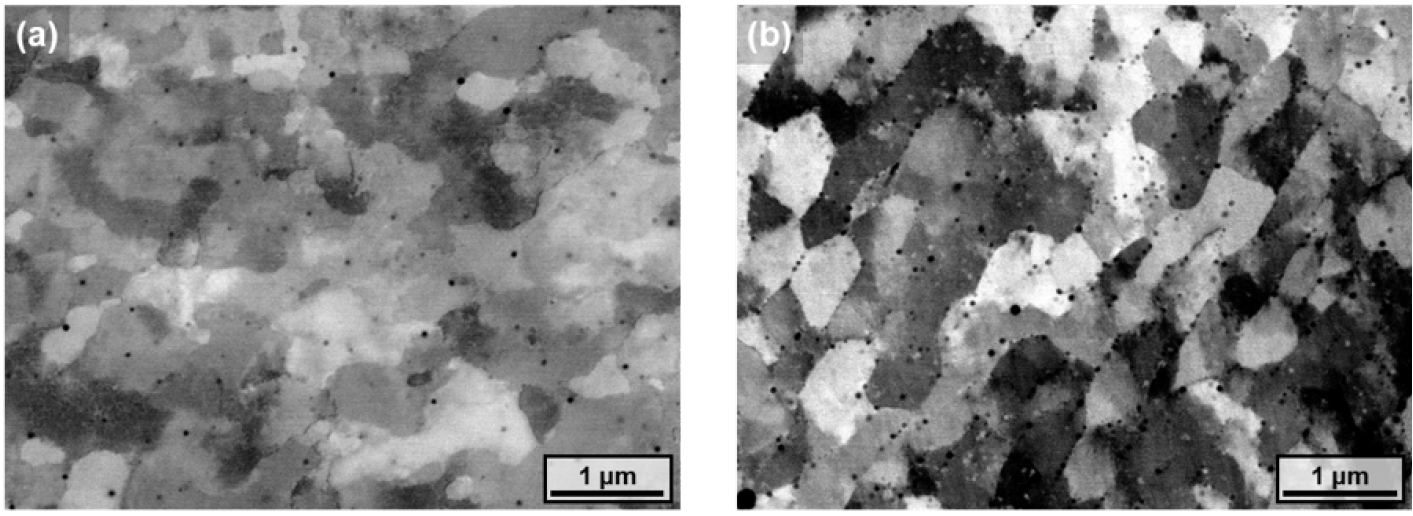
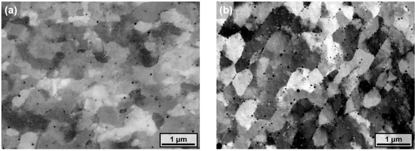
- Figure 5 shows the BSE microstructures of the two samples. Fine dark-contrast particles, interpreted as oxide particles, were more frequently observed in the Oxidized sample. Image analysis of the BSE fields shown in Fig. 5 yielded approximate mean equivalent circular diameters of 51 ± 8 and 50 ± 13 nm for the segmented oxide particles in the Virgin and Oxidized samples, respectively. Their corresponding apparent area fractions were 0.37% and 1.81%, indicating similar particle sizes but a higher area fraction in the Oxidized sample. Dispersed oxide particles have been shown to impede dislocation motion in Cu [20]. Although the exact oxide phase (Cu2O or CuO) was not directly identified, the observed particle distribution suggests a possible contribution to strengthening. The particle-strengthening contribution (Δσp) is discussed qualitatively because it was not independently quantified. The higher strength of the Oxidized sample may thus reflect both particle strengthening and a greater dislocation-strengthening contribution, consistent with the BSE and KAM observations. The comparable relative densities and limited difference in grain size suggest that densification and grain refinement are unlikely to be the primary causes of the strength increase.
3. Results and discussion
- In this study, virgin and oxidized pure Cu powders were processed under identical LPBF conditions, and the relative density, tensile properties, and microstructural characteristics of the resulting samples were compared. The main conclusions are as follows:
- (1) The Virgin and Oxidized samples exhibited comparable relative densities of 98.8% and 98.7%, respectively, indicating that both powder conditions produced similar densification under the applied LPBF condition.
- (2) The YS and UTS of the Oxidized sample were 195 and 239 MPa, corresponding to increases of 24.2% and 13.3%, respectively, compared with the Virgin sample. These increases were accompanied by decreases in uniform and total elongation.
- (3) The grain sizes of the two samples were similar, whereas the mean KAM value increased from 1.49° in the Virgin sample to 2.18° in the Oxidized sample. Image analysis of the examined BSE fields indicated similar mean particle sizes but a higher apparent area fraction in the Oxidized sample. The enhanced strength of the Oxidized sample may therefore be associated with both oxide-particle strengthening and a greater dislocation-strengthening contribution. The oxide particles may also promote dislocation accumulation and retention, providing a possible link between the particle distribution and the higher KAM value.
4. Conclusions
-
Funding
This work was supported by Fundamental R&D Projects (PNKB140 and PNKB310) of the Korea Institute of Materials Science (KIMS). This work was also supported by the National Research Foundation of the Republic of Korea (NRF) [grant number: RS-2023-00282305] and the Ministry of Trade, Industry and Energy of the Republic of Korea (MOTIE) [grant number: RS-2025-02317513].
-
Conflict of Interest
The authors declare that they have no conflict of interest.
-
Data Availability Statement
The data that support the findings of this study are not publicly available due to confidentiality agreements with industrial partners.
-
Author Information and Contribution
Haeum Park: Data Curation, Formal analysis, Investigation, Methodology, Validation, Visualization, Writing – original draft
Ki Jong Kim: Data Curation, Investigation, Methodology, Writing – original draft
Yeon Woo Kim: Data Curation, Investigation, Methodology
Hahun Lee: Data Curation, Formal analysis, Validation
Kichul Park: Methodology, Resources, Validation
Yerim Kim: Data Curation, Investigation, Validation
Kee-Ahn Lee: Validation
Seok Su Sohn: Methodology, Resources
Jeong Min Park: Conceptualization, Data curation, Investigation, Methodology, Project administration, Supervision, Validation, Visualization, Writing – review & editing
-
Acknowledgments
None.
Article information
| Sample | YS (MPa) | UTS (MPa) | T. El. (%) | U. El. (%) |
|---|---|---|---|---|
| Virgin | 157 | 211 | 38.8 | 27.4 |
| Oxidized | 195 | 239 | 28.6 | 19.6 |
- 1. M. Abdelhafiz, A. Emadi and M. A. Elbestawi: Materials, 16 (2023) 4694.Article
- 2. J. Robinson, S. P. Munagala, A. Arjunan, N. Simpson, R. Jones, A. Baroutaji, L. T. Govindaraman and I. Lyall: Materials, 15 (2022) 7563.Article
- 3. S. A. Niknam, M. Mortazavi and D. Li: Int. J. Adv. Manuf. Technol., 112 (2021) 601.ArticlePDF
- 4. Q. Jiang, P. Zhang, Z. Yu, H. Shi, D. Wu, H. Yan, X. Ye, Q. Lu and Y. Tian: Coatings, 11 (2021) 740.Article
- 5. M. Roccetti Campagnoli, M. Galati and A. Saboori: J. Manuf. Process., 72 (2021) 320.Article
- 6. M. Colopi, A. G. Demir, L. Caprio and B. Previtali: Int. J. Adv. Manuf. Technol., 104 (2019) 2473.ArticlePDF
- 7. T.-T. Ikeshoji, K. Nakamura, M. Yonehara, K. Imai and H. Kyogoku: JOM, 70 (2018) 396.ArticlePDF
- 8. M. Colopi, L. Caprio, A. G. Demir and B. Previtali: Procedia CIRP, 74 (2018) 59.Article
- 9. S. D. Jadhav, S. Dadbakhsh, L. Goossens, J.-P. Kruth, J. Van Humbeeck and K. Vanmeensel: J. Mater. Process. Technol., 270 (2019) 47.Article
- 10. C. Silbernagel, L. Gargalis, I. Ashcroft, R. Hague, M. Galea and P. Dickens: Addit. Manuf., 29 (2019) 100831.Article
- 11. S. Gruber, L. Stepien, E. López, F. Brueckner and C. Leyens: Materials, 14 (2021) 3642.Article
- 12. E. Hori, Y. Sato, T. Shibata, K. Tojo and M. Tsukamoto: J. Laser Appl., 33 (2021) 012008.Article
- 13. G. Nordet, C. Gorny, Y. Mayi, J. Daligault, M. Dal, A. Effernelli, E. Blanchet, F. Coste and P. Peyre: Opt. Laser Technol., 147 (2022) 107612.Article
- 14. M. S. Khandpur, A. Giubilini, L. Iuliano and P. Minetola: Metals, 14 (2024) 1464.Article
- 15. S. D. Jadhav, S. Dadbakhsh, J. Vleugels, J. Hofkens, P. Van Puyvelde, S. Yang, J.-P. Kruth, J. Van Humbeeck and K. Vanmeensel: Materials, 12 (2019) 2469.Article
- 16. V. Lindström, O. Liashenko, K. Zweiacker, S. Derevianko, V. Morozovych, Y. Lyashenko and C. Leinenbach: Materials, 13 (2020) 3493.Article
- 17. S. D. Jadhav, J. Vleugels, J.-P. Kruth, J. Van Humbeeck and K. Vanmeensel: Mater. Des. Process. Commun., 2 (2020) e94.Article
- 18. R. N. Gu, P. Chen, Y. H. Zhou, H. Wang, X. C. Yan, K. S. Wong and M. Yan: Adv. Eng. Mater., 25 (2023) 2101138.Article
- 19. Y. Liu, J. Zhang, R. Niu, M. Bayat, Y. Zhou, Y. Yin, Q. Tan, S. Liu, J. H. Hattel, M. Li, X. Huang, J. Cairney, Y.-S. Chen, M. Easton, C. Hutchinson and M.-X. Zhang: Nat. Commun., 15 (2024) 1283.Article
- 20. S. Qu, L. Wang, S. Zhang, C. Yang, H. Y. Chia, G. Wu, Z. Hu, J. Ding, W. Yan, Y. Zhang, C. H. Chan, W. Chen, Y. Lu and X. Song: Nat. Commun., 16 (2025) 3234.Article
- 21. A. Kolhatkar, V. Karthik, G. M. S. K. Chaitanya, A. Kumar and D. Ramchandran: J. Test. Eval., 47 (2019) 3417.ArticlePDF
- 22. M. Mandal, B. Aashranth, A. Kolhatkar, M. A. Davinci, D. Samantaray, V. Karthik and M. Vasudevan: J. Mater. Sci.: Mater. Eng., 20 (2025) 54.Article
- 23. J. I. Yoon, J. G. Kim, J. M. Jung, D. J. Lee, H. J. Jeong, M. Shahbaz, S. Lee and H. S. Kim: Korean J. Met. Mater., 54 (2016) 231.Article
- 24. ASTM International, ASTM E8/E8M-24, Standard Test Methods for Tension Testing of Metallic Materials, ASTM International, West Conshohocken, PA (2024).
- 25. H. Park, H. Kwon, K. T. Kim, J.-H. Yu, J. Choe, H. Sung, H. S. Kim, J. G. Kim and J. M. Park: Addit. Manuf., 86 (2024) 104223.Article
- 26. J. M. Park, E. S. Kim, H. Kwon, P. Sathiyamoorthi, K. T. Kim, J.-H. Yu and H. S. Kim: Addit. Manuf., 47 (2021) 102283.Article
- 27. Y. S. Eom, J. M. Park, J.-W. Choi, D.-J. Seong, H. Joo, Y. C. Jo, K. T. Kim, J.-H. Yu and I. Son: J. Alloys Compd., 956 (2023) 170348.Article
- 28. K. M. Bertsch, G. Meric de Bellefon, B. Kuehl and D. J. Thoma: Acta Mater., 199 (2020) 19.Article
- 29. L. Gao, Y. Chen, X. Zhang, S. R. Agnew, A. C. Chuang and T. Sun: Nat. Commun., 16 (2025) 4696.Article
References
Figure & Data
References
Citations

-
 Cite this Article
- Cite this Article
-
- Close
- Download Citation
- Close
 XML Download
- Figure
-
- Related articles
-
- Microstructure, Magnetic Properties, and Performance of Fe-6.5Si Soft Magnetic Core Produced by Laser Powder Bed Fusion
- Effect of Building Orientation on Tensile Properties of Hastelloy X alloy Manufactured by Laser Powder Bed Fusion
- Ultra-Low-Temperature (4.2 K) Tensile Properties and Deformation Mechanism of Stainless Steel 304L Manufactured by Laser Powder Bed Fusion
- Effect of Hatch Spacing on the Microstructure and Mechanical Properties of SA508 Gr.3 Steel Fabricated by Laser Powder Bed Fusion
- Effect of Bulk Shape on Mechanical Properties of Ti-6Al-4V Alloy Manufactured by Laser Powder Bed Fusion
Fig. 1.
Fig. 2.
Fig. 3.
Fig. 4.
Fig. 5.
Graphical abstract
| Sample | YS (MPa) | UTS (MPa) | T. El. (%) | U. El. (%) |
|---|---|---|---|---|
| Virgin | 157 | 211 | 38.8 | 27.4 |
| Oxidized | 195 | 239 | 28.6 | 19.6 |
Table 1.
TOP

